
Midfoot Arthritis: What Your Foot X-ray Shows and When to Scan Further
Midfoot arthritis is wear of the cartilage in the joints across the middle of the foot, often the tarsometatarsal joints at the bases of the first three metatarsals. A standing X-ray is the first test and usually enough: it shows narrowed joint spaces, hardened bone edges and a spur on top of the foot. CT, MRI or a guided injection is added only for specific questions.
Common Symptoms
- Aching across the top and middle of the foot that is worse when standing, walking uphill or climbing stairs
- A hard, bony bump on the top of the midfoot that rubs against shoe laces or the tongue of a shoe
- Stiffness in the middle of the foot when getting up in the morning or after sitting
- Pain when pushing off through the ball of the foot or rising onto the toes
- Tingling or numbness between the big toe and second toe where the bump presses on a nerve
- An arch that slowly drops or a foot that turns outward over months or years
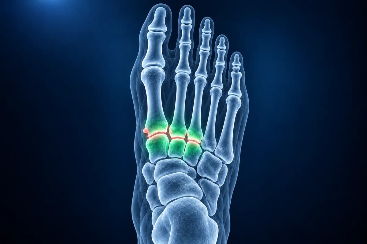
The midfoot is a block of small bones that turns the foot into a rigid lever when you push off. When the cartilage between those bones thins, the joints grind and form bony lips, and the arch can slowly sag. A review by Patel and colleagues names an old injury as the leading cause, but age-related wear, an inflammatory arthritis or nerve damage from diabetes can lead to the same picture. This page explains the imaging, the phrases your report may use and the questions worth asking. It is educational guidance, not a diagnosis.
Which joints does midfoot arthritis affect?
The middle of the foot holds three rows of joints. The tarsometatarsal joints, also called the Lisfranc joints, join the five long metatarsals to the cuneiforms and the cuboid. Behind them sit the joints between the navicular and the cuneiforms, and the talonavicular joint at the back of the arch. The inner tarsometatarsal joints form a stiff column that carries load during push-off and normally move only a few degrees. The fourth and fifth tarsometatarsal joints move much more, which helps the outer foot adapt to uneven ground.
In a community study of adults aged 50 and over in England, Roddy and colleagues estimated that roughly one in six people had painful foot osteoarthritis visible on X-ray, and of the five joints they graded, the second cuneiform–metatarsal joint was the most often involved after the big toe joint. If the big toe joint is your main problem, the hallux rigidus page covers it; both forms of arthritis often show on the same X-ray.
Why does midfoot arthritis cause a bump on top of the foot?
As the joint wears, the bone at its upper edge grows a lip called a dorsal osteophyte. Because the top of the midfoot has almost no padding, this spur is easy to feel and presses against laces, straps and stiff shoe tongues. The deep peroneal nerve runs right over it, so some people notice tingling or numbness in the web space between the big toe and the second toe.
A bump on top of the foot is not always bone. A soft, slightly mobile swelling that changes size is more often a ganglion cyst, which can grow out of an arthritic joint. The guide to top of foot pain and lumps compares the common causes and the test that sorts each one out.
Which imaging test comes first for midfoot arthritis?
For chronic foot pain, the American College of Radiology Appropriateness Criteria rate plain X-rays as the first test. For the midfoot, the views should be taken standing: weight on the foot narrows a worn joint and opens up any sag of the arch that a lying-down film can hide. A front view, a side view and an oblique view are standard. The oblique view matters because it shows the third and fourth tarsometatarsal joints without the bones overlapping. If you already have the images, you can open them in the free X-ray viewer.
CT shows the joint surfaces, cysts and spurs in fine detail. It is mainly used to plan surgery or when the X-ray is unclear. Weight-bearing CT, where it is available, shows the joints under load in three dimensions; the weight-bearing CT explainer covers what it adds.
MRI is useful when the X-ray looks nearly normal but pain persists. It shows bone marrow edema next to an inflamed joint, an early stress fracture such as a navicular stress fracture, tendon problems and ligament damage. Ultrasound is good for a lump on top of the foot and for guiding a needle. When several joints look worn, an image-guided injection of local anesthetic into one joint at a time can show which joint is actually causing the pain, which matters before a fusion.
What does “degenerative change at the tarsometatarsal joints” mean on your report?
These are the phrases midfoot X-ray, CT and MRI reports use most often. The foot X-ray report phrases guide explains the wider vocabulary.
- “Joint space narrowing at the second and third tarsometatarsal joints”: the gap that holds cartilage is thinner than it should be. Ask whether the narrowing matches where you feel the pain.
- “Dorsal osteophytes” or “dorsal spurring”: bony lips on the top of the joint, the bump you can feel. Ask whether the spur could be pressing on the nerve if you have numbness near the big toe.
- “Subchondral sclerosis” and “subchondral cysts”: the bone under the cartilage has become denser or has small fluid pockets, both signs of longstanding wear. Ask how advanced the arthritis is overall.
- “Midfoot sag” or “plantar gapping at the first tarsometatarsal joint”: on the standing side view, the arch bends at the midfoot instead of at the ankle. Ask whether it changes shoe or insole advice; the foot X-ray angles guide shows how the arch is measured.
- “Subchondral bone marrow edema at the tarsometatarsal joints” on MRI: fluid in the bone on both sides of a worn joint, a sign that the arthritis is currently active. Ask whether it explains a recent flare.
- “Fragmentation” or “joint disorganization”: broken-up bone and debris around the joints. In someone with diabetes or reduced feeling in the foot, ask directly whether this could be Charcot foot, because the treatment is different and urgent.
What We Look For on Midfoot Imaging
- Joint space width at each tarsometatarsal and naviculocuneiform joint on standing views
- Dorsal osteophytes, subchondral sclerosis and subchondral cysts at the bases of the metatarsals
- Alignment of the first and second metatarsal bases, including any widening or step-off
- Sag of the arch at the midfoot on the standing side view
- Bone marrow edema, stress fracture lines or fragmentation on MRI
- Soft-tissue lumps over the joints, such as a ganglion arising from the joint capsule
Is it wear and tear, or an old Lisfranc injury?
Arthritis in one or two midfoot joints in a younger adult, especially after a twist, a fall from a step or a sports injury, raises the question of an old Lisfranc injury. A ligament tear between the first cuneiform and the second metatarsal can heal with the bones slightly apart, and that small shift wears the joint out years later. Clues on the X-ray include a small bone fleck between the first two metatarsals and a step between the second metatarsal and its cuneiform. The article on Lisfranc injuries missed on X-ray explains why these injuries are overlooked at first.
Arthritis that involves many joints in both feet, with swelling and long-lasting morning stiffness, points instead toward an inflammatory arthritis such as rheumatoid arthritis of the foot, which needs blood tests and a rheumatology opinion rather than shoe changes alone.
When should midfoot pain be checked urgently?
- You have diabetes or numb feet and the midfoot becomes warm, red or swollen, even without pain. Same-day assessment is needed to rule out an active Charcot foot or infection.
- You cannot put weight on the foot after an injury, or there is bruising on the sole of the midfoot.
- Fever, a hot joint, an open wound or pus near the painful area.
- Pain that wakes you at night, comes with unexplained weight loss or does not change with rest or activity.
How is midfoot arthritis treated, and when is fusion considered?
Most treatment aims to stop the worn joints from bending. Stiff-soled shoes, a rocker sole, a carbon-fiber insole or a custom orthosis that supports the arch reduce motion through the midfoot during push-off. Lacing that skips the eyelets over the bump, pain relief and adjusting high-impact activity help many people. A steroid injection placed with ultrasound or X-ray guidance can calm a flare and, at the same time, confirms which joint is responsible.
When pain stays despite these measures, surgeons usually offer a fusion (arthrodesis) of the painful inner-column joints, joining the bones so they no longer grind. Because the first three tarsometatarsal joints normally move very little, fusing them costs little function. The fourth and fifth joints are usually spared where possible because the outer foot needs their motion, and how best to treat them is still debated. Removing a spur alone can help when the bump is the main problem.
Frequently Asked Questions
Can midfoot arthritis be seen on an X-ray?
Usually, yes. Standing X-rays show narrowed joint spaces, spurs on top of the foot, denser bone and small cysts under the cartilage. Early arthritis can look normal on X-ray, and MRI or CT may be added if the pain does not match what the X-ray shows.
Why does my X-ray need to be taken standing?
Weight squeezes the joints and lets the arch settle into its true position. A worn joint can look wider and the arch higher on an X-ray taken lying down, so standing views give a more honest picture of joint narrowing, arch sag and any old ligament injury.
Is the bony bump on top of my foot dangerous?
A hard bump over a worn midfoot joint is usually a bone spur, which is harmless but can be sore in shoes and irritate a nerve. A lump that is growing, soft and changing in size, or painful at night should be examined and may need an ultrasound to tell a spur from a cyst or another kind of lump.
Do I need an MRI for midfoot arthritis?
Often not. When the standing X-ray clearly shows arthritis that matches your pain, an MRI rarely changes the plan. MRI helps when the X-ray is near normal, when a stress fracture or tendon problem is suspected, or when several joints look worn and the surgeon needs to know which one is active.
Can AI read my midfoot X-ray report?
Weight-bearing X-rays show joint-space narrowing, spurs and alignment, but the painful joint must be identified clinically. AI cannot determine which joint needs treatment.
Sources
- Expert Panel on Musculoskeletal Imaging; Said N, Fox MG, Nacey NC, et al. ACR Appropriateness Criteria Chronic Foot Pain: Update 2025. J Am Coll Radiol. 2026;23(4):681-699.
- Patel A, Rao S, Nawoczenski D, Flemister AS, DiGiovanni B, Baumhauer JF. Midfoot arthritis. J Am Acad Orthop Surg. 2010;18(7):417-425.
- Roddy E, Thomas MJ, Marshall M, et al. The population prevalence of symptomatic radiographic foot osteoarthritis in community-dwelling older adults: cross-sectional findings from the clinical assessment study of the foot. Ann Rheum Dis. 2015;74(1):156-163.
- Menz HB, Munteanu SE, Landorf KB, Zammit GV, Cicuttini FM. Radiographic classification of osteoarthritis in commonly affected joints of the foot. Osteoarthritis Cartilage. 2007;15(11):1333-1338.
- American Academy of Orthopaedic Surgeons. Arthritis of the Foot and Ankle. OrthoInfo.
Understand your imaging before your next appointment
MRI, X-ray or CT from a CD, folder or ZIP. A PDF report or photos of the films work too.
Free to open and preview, no account needed. An optional AI report from $10 explains your images in plain language, with questions for your doctor.
Your raw files stay on this device. Only rendered images and minimal context are sent when you request an AI report.
Medical Disclaimer: This page is for informational and educational purposes only. It does not constitute medical advice, diagnosis, or treatment. AI-generated analysis may contain errors. Always consult a qualified healthcare professional for medical decisions. Full Disclaimer