
Jones Fracture: Fifth Metatarsal Zones, Avulsion Look-Alikes and Healing
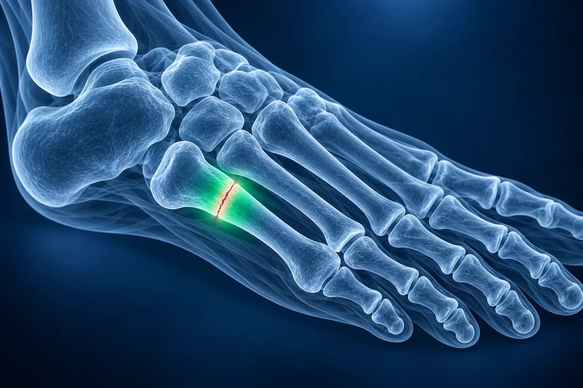
A Jones fracture is a break in the fifth metatarsal about 1.5 to 2 cm beyond its base, at the junction where the wide base meets the shaft (zone 2). It differs from the more common avulsion fracture at the very tip of the base (zone 1) because blood supply there is poor, so it heals more slowly and is more likely to need a screw.
Common Symptoms
- Pain and tenderness along the outer edge of the foot, a thumb-width beyond the bony bump at the fifth metatarsal base
- Swelling and bruising on the outside of the midfoot after a twist, cut or landing
- Pain when walking, especially when pushing off or turning on the forefoot
- An ache on the outside of the foot for weeks before a sudden worsening, which suggests a stress fracture
- Pain when standing on the outer border of the foot or rising onto the toes
When is an outer foot injury urgent?
Seek emergency care if you could not take four steps right after the injury or still cannot, if the foot looks deformed, if skin is broken over the bone, or if the toes become numb, pale or cold. After a crush or high-energy injury, pain that keeps climbing with a tight, swollen foot and pain on stretching the toes can signal compartment syndrome. People with diabetes and reduced feeling in the feet should be seen quickly for any new swelling.
Is it a Jones fracture or an avulsion fracture?
Radiologists divide the proximal fifth metatarsal into three zones, a scheme popularised by Lawrence and Botte. Where the line sits on the X-ray matters more than how dramatic it looks.
- Zone 1: tuberosity avulsion ("pseudo-Jones")
- A fragment pulled off the very end of the base, typically when the foot rolls inward. It is by far the most common fifth metatarsal base fracture and usually heals well in a stiff shoe or walking boot.
- Zone 2: the true Jones fracture
- A roughly transverse line at the junction of the base and shaft, aimed at the joint between the fourth and fifth metatarsals. Blood vessels reach this segment from opposite directions, leaving a watershed area that is slow to heal.
- Zone 3: proximal shaft stress fracture
- A fracture slightly further along the shaft, usually from repeated loading in athletes. It often shows thickened cortex or a widened gap on X-ray and carries the highest risk of delayed union.
A spiral break further toward the toes, sometimes called a dancer's fracture, is a different injury of the distal shaft. For more on the overlap between gradual and sudden breaks, see our metatarsal stress fracture page.
How does a Jones fracture happen?
The typical mechanism is a large force through the outer forefoot while the heel is off the ground: a basketball player planting and cutting, a footballer changing direction, or a dancer landing on the outer foot. Some patients recall an ache along the outside of the foot for weeks, meaning a stress injury was already developing before the final break. A high-arched foot that loads its outer border, and the same bone-health factors that drive other stress fractures, can contribute. The same rolling movement also sprains the outer ankle ligaments, so the two injuries are often examined together, but the fifth metatarsal is part of the foot exam.
Pain that sits further toward the middle of the foot after a twist is more typical of a Lisfranc injury, and a bony, aching outer foot in a teenager with stiff, recurrent sprains can come from a tarsal coalition. Those are worth raising if your X-ray of the fifth metatarsal is normal. Explore midfoot pain patterns when the pain is not centred on the fifth metatarsal.
Do I need an X-ray, and what does it show?
The Ottawa foot rules recommend foot X-rays after an injury when there is midfoot pain plus bone tenderness at the base of the fifth metatarsal or at the navicular, or when you could not bear weight for four steps both immediately and when examined. The ACR Appropriateness Criteria for acute foot trauma support three standard views (front, oblique and side) as the first test. The oblique view usually shows the fifth metatarsal base best.
CT is helpful when healing is uncertain on X-ray, for example to decide whether a gap has bridged months after treatment. MRI is used when pain on the outside of the foot persists with normal X-rays, to look for an early stress reaction in zone 3 or for problems in the peroneal tendons that attach nearby.
What do fifth metatarsal report phrases mean?
- "Fracture at the metaphyseal-diaphyseal junction"
- The break is where the wide base narrows into the shaft. This is the location that defines a true Jones fracture.
- "Extends into the fourth-fifth intermetatarsal articulation"
- The crack runs toward the small joint between the bases of the fourth and fifth metatarsals, another marker of zone 2.
- "Avulsion fracture of the fifth metatarsal tuberosity"
- A zone 1 chip from the tip of the base, the more forgiving injury.
- "Intramedullary sclerosis" or "cortical thickening"
- Dense bone inside or around the fracture, a sign the injury has been building over time and a risk factor for slow healing.
- "Unfused apophysis" or "os peroneum"
- Normal variants. In children and teenagers a growth plate runs along the base, roughly parallel to the shaft; a fracture usually runs across it. Small extra bones near the base can also look like fragments.
To practise reading these lines on your own films, see how to read a foot X-ray.
What we look for on fifth metatarsal imaging
- Fracture position relative to the tuberosity tip and the fourth-fifth intermetatarsal joint
- Fracture direction: transverse across the bone versus a growth plate running along it
- Gap width, displacement and whether the line reaches the joint surface
- Sclerotic margins or cortical thickening that suggest a chronic stress fracture
- Bridging callus or a persistent lucent gap on follow-up X-ray or CT
Does a Jones fracture need surgery?
Not always. A non-displaced acute zone 2 fracture can heal in a cast or boot without weight on the foot, usually for about six to eight weeks, followed by protected walking. Healing is slower than for most foot fractures, and some go on to delayed union or nonunion. Many surgeons therefore offer an intramedullary screw to competitive athletes, to people with zone 3 or chronic fractures, and to anyone whose fracture has not healed with a cast. The decision balances the risks of surgery against time off your feet, and your surgeon will weigh both with you.
Zone 1 avulsion fractures, by contrast, usually heal in a supportive shoe or boot with weight bearing as comfort allows over several weeks. Our metatarsal fracture healing timeline compares the expected course of each type.
Can I review my own fifth metatarsal X-ray?
Open your DICOM files privately in the free foot X-ray viewer to see where the line sits relative to the base. For a plain-language explanation of the zone, healing signs and report wording, upload your imaging for an AI review. It explains what is visible; it does not diagnose, and treatment decisions stay with your clinician. Use the foot X-ray report guide alongside the Jones zone description.
Frequently Asked Questions
Can you walk on a Jones fracture?
Many people can limp on a Jones fracture, but walking on it before it heals raises the risk of the bone not joining. Non-operative treatment usually means no weight on the foot for several weeks. Follow the plan your clinician gives you, which depends on the zone and whether the fracture was treated with a screw.
Why does a Jones fracture heal so slowly?
The segment where a zone 2 fracture occurs sits between two blood supplies, one entering the shaft and one supplying the base, so it receives less blood than surrounding bone. Forces from the tendons that pull on the base also keep moving the fracture. Smoking and stress-related bone changes slow healing further.
How long is recovery after a Jones fracture screw?
Timelines vary by surgeon and patient, but recovery is usually measured in weeks to a few months, with a period of protected walking before running. Return to sport is generally based on pain-free activity and evidence of healing on X-ray or CT rather than a fixed date.
My child's X-ray shows a line at the fifth metatarsal base. Is it a fracture?
Possibly not. Children and young teenagers have a growth plate (apophysis) at the base that appears as a thin line running along the length of the bone. Fractures usually run across the bone instead. If the area is tender, a clinician compares the X-ray with the examination and sometimes with the other foot.
Sources
- Lawrence SJ, Botte MJ. Jones' fractures and related fractures of the proximal fifth metatarsal. Foot Ankle. 1993;14(6):358-365.
- Smidt KP, Massey P. 5th Metatarsal Fracture. StatPearls. Treasure Island (FL): StatPearls Publishing.
- Stiell IG, Greenberg GH, McKnight RD, et al. A study to develop clinical decision rules for the use of radiography in acute ankle injuries. Ann Emerg Med. 1992;21(4):384-390.
- Expert Panel on Musculoskeletal Imaging; Gorbachova T, Chang EY, Ha AS, et al. ACR Appropriateness Criteria Acute Trauma to the Foot. J Am Coll Radiol. 2020;17(5S):S2-S11.
- American Academy of Orthopaedic Surgeons. Toe and Forefoot Fractures. OrthoInfo.
Related Articles
How long a broken metatarsal takes to heal week by week, why the fracture line lingers on X-ray, which fractures heal slowly, and what follow-up reports mean.
Why a foot stress fracture can hide on a normal X-ray, what MRI, CT and repeat X-rays show instead, and which foot stress fractures are high risk.
What hallux valgus angle, fleck sign, joint space narrowing at the first MTP, pes planus, calcaneal spur and Jones fracture mean on your foot X-ray report.
Understand your imaging before your next appointment
MRI, X-ray or CT from a CD, folder or ZIP. A PDF report or photos of the films work too.
Free to open and preview, no account needed. An optional AI report from $10 explains your images in plain language, with questions for your doctor.
Your raw files stay on this device. Only rendered images and minimal context are sent when you request an AI report.
Medical Disclaimer: This page is for informational and educational purposes only. It does not constitute medical advice, diagnosis, or treatment. AI-generated analysis may contain errors. Always consult a qualified healthcare professional for medical decisions. Full Disclaimer