
Stress Fracture Not Showing on X-Ray: Why and What Comes Next
Why a foot stress fracture can hide on a normal X-ray, what MRI, CT and repeat X-rays show instead, and which foot stress fractures are high risk.
Key Takeaways
- A normal X-ray in the first weeks does not rule out a stress fracture
- MRI shows bone marrow edema and fracture lines before X-rays change and is the usual next test
- CT helps with the navicular and surgical planning; a repeat X-ray suits low-risk sites
- Navicular, fifth metatarsal base, sesamoid and second metatarsal base stress fractures are high risk
- A warm swollen foot in diabetes with a normal X-ray may be early Charcot foot and needs prompt care
- Many foot stress fractures heal in around 6 to 8 weeks; high-risk sites often take longer

Yes, you can have a stress fracture with a normal X-ray. In the first weeks the crack is too fine to see, and the American Academy of Orthopaedic Surgeons notes the line may only appear several weeks later, once healing bone has formed. If a stress fracture is still suspected, the American College of Radiology recommends MRI as the usual next test; a repeat X-ray after a couple of weeks or a CT scan are alternatives in some situations.
A normal first X-ray is therefore not the end of the story. It rules out a displaced fracture and other bone problems, but it cannot rule out an early stress injury. This article explains why, which tests see what the X-ray cannot, how to read the report phrases that follow, and which foot stress fractures need extra caution. It is educational information and not a diagnosis for your own foot.
Why doesn't a stress fracture show on the first X-ray?
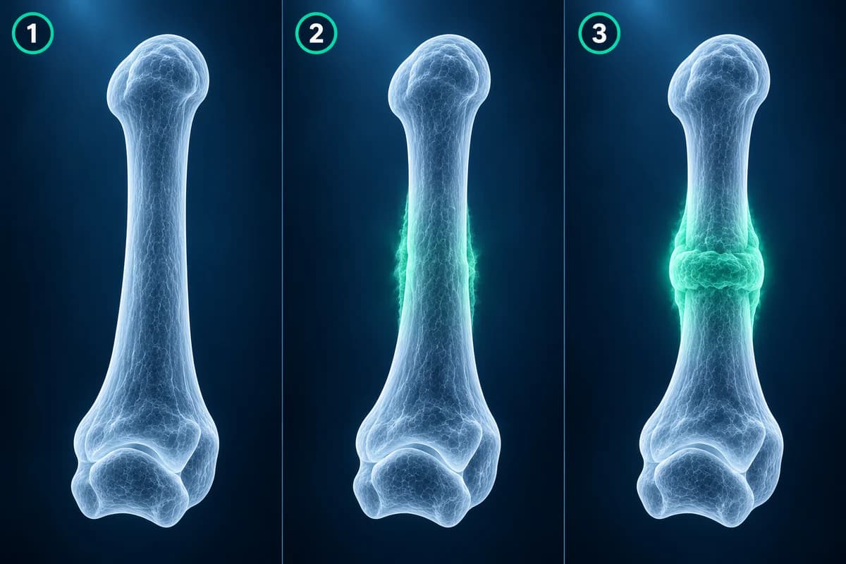
A stress fracture is not a single event like a twisted ankle. It develops when bone is loaded faster than it can repair itself, typically after a jump in running distance, a new sport, a change of footwear, or long marches. The earliest change is inside the bone: tiny cracks in the internal scaffolding and fluid in the marrow. None of that changes how much X-ray the bone absorbs, so the image looks normal.
X-rays become positive when the body starts to heal. New bone forms along the surface of a metatarsal as a faint, fluffy cuff, or a dense white band appears across the heel bone. By the time those signs are visible the injury has often been present for weeks. Some stress fractures never show clearly on plain films at all, particularly in the navicular, a small midfoot bone whose shape and overlap with its neighbors hide fine lines.
Which test shows a stress fracture the X-ray missed?
MRI is the most useful next step for most people. Fluid-sensitive sequences light up the marrow swelling of an early stress injury days to weeks before an X-ray changes, and they can show the fracture line itself as a dark stripe. MRI also shows what else could be causing the pain, from a neuroma to a plantar plate tear, without radiation. The 2024 ACR criteria on stress fractures rate it the preferred study when X-rays are negative or indeterminate.
CT is better at showing the exact path of a fracture line through dense bone and is often used for the navicular, or when a surgeon needs to see whether a crack runs all the way across. A bone scan detects increased bone turnover and is sensitive, but less specific and it involves radiation, so it is used less often now. A repeat X-ray after about two weeks costs little and may show healing changes, which is a reasonable route when the suspected fracture is in a low-risk spot.
If you already have an MRI, the free MRI viewer opens the study privately in your browser. For a refresher on the sequences, see how to read a foot MRI.
What does "bone marrow edema" mean on a foot MRI?
"Bone marrow edema in the second metatarsal" means extra fluid inside that bone, seen as bright signal on fluid-sensitive images. In a runner with focal pain, it usually reflects stress. On its own it is not a diagnosis: bruising, arthritis, infection and Charcot foot can also cause it, so it is always read alongside the symptoms.
"Stress reaction" is the term for marrow and surface swelling without a visible fracture line, the early end of the spectrum. "Periosteal edema" is fluid along the thin membrane that wraps the bone, often the first sign. "Stress fracture" or "fracture line" means a dark line can be seen crossing part or all of the bone. Some reports grade the injury from mild to severe based on how far along this sequence it is, which helps estimate how long protection is needed.
On X-ray reports, "periosteal reaction" or "callus formation" means healing bone is forming around a fracture, and "sclerotic band" means a dense white line across the bone, typical of a healing heel bone stress fracture.
Which foot stress fractures are high risk?
Location matters more than size. The shafts of the second and third metatarsals are the classic low-risk sites; a metatarsal stress fracture there usually heals with reduced load and a stiff shoe or boot. The heel bone is another common and usually forgiving site.
High-risk sites have poor blood supply or high tension, and are more likely to heal slowly, fail to heal, or progress to a complete break. In the foot these are the navicular, the base of the fifth metatarsal just beyond the flare where a Jones fracture occurs, the sesamoid bones under the big toe joint, and the base of the second metatarsal. A suspected stress injury at any of these deserves early imaging rather than a wait-and-see repeat X-ray.
What else can look like a stress fracture?
Pain under the ball of the foot with a normal X-ray has several causes. A plantar plate tear hurts under a toe joint, and the toe may start to drift. A Morton's neuroma causes burning between the toes. Pain under the big toe joint can be sesamoiditis or a sesamoid stress fracture, which can be hard to tell apart without MRI or CT. In teenagers, Freiberg disease affects the head of the second metatarsal and can start with a normal X-ray.
One mimic matters above the rest. In a person with diabetes and reduced feeling in the feet, a warm, swollen foot with marrow swelling and a normal X-ray can be the earliest stage of Charcot foot. Catching it before the bones collapse depends on prompt offloading, so this combination should be seen by a clinician quickly, not managed as a simple overuse injury.
What should you do if the X-ray is normal but the pain persists?
Tell your clinician that the pain is still there and where exactly it is. Pinpoint tenderness over one bone, pain that builds during activity and lingers afterward, and swelling on the top of the foot are the features that keep a stress fracture on the table. Until it is ruled out, clinicians commonly advise reducing impact, and sometimes a stiff-soled shoe or walking boot.
Once confirmed, many foot stress fractures take around 6 to 8 weeks to heal according to the AAOS, with high-risk sites often taking longer. Our metatarsal fracture healing timeline covers the stages. Repeated stress fractures are a reason to ask about bone health, nutrition and training load, not only about the foot. You can also upload your images for an educational read of what the X-ray or MRI shows.
When is foot pain an emergency?
Seek urgent care if you cannot bear weight at all, if the foot is badly swollen or deformed after an injury, if toes become pale, cold or numb, if you have a fever, or if you have diabetes and the foot becomes hot, red or swollen. Those situations need examination the same day, whatever an earlier X-ray showed.
Frequently Asked Questions
How long after a stress fracture does it show on X-ray?
Often several weeks, because the X-ray usually shows the healing response rather than the crack itself. Some stress fractures, especially in the navicular and sesamoids, may never be clearly visible on plain films. That is why MRI or CT is used when the suspicion remains.
Can I walk on a stress fracture that does not show on X-ray?
Continuing to load a stress injury can turn a crack into a complete fracture. Until a clinician has assessed it, it is sensible to reduce impact and avoid running or jumping on the painful foot. Whether walking is allowed, and with what shoe or boot, depends on the bone involved and should be decided with your clinician.
Is a stress reaction the same as a stress fracture?
They are points on the same spectrum. A stress reaction means swelling in and around the bone without a visible fracture line. A stress fracture means a line can be seen. A stress reaction is earlier and usually heals faster, but it can progress if loading continues.
Why did my doctor order a CT instead of an MRI?
CT shows fine cortical detail and the exact course of a fracture line, which matters for the navicular and for deciding whether surgery is needed. MRI is better at early detection and at showing soft tissues. The choice depends on the bone involved, what has already been done, and whether MRI is available or suitable for you.
Can bone marrow edema be something other than a stress fracture?
Yes. Bone bruising after an injury, arthritis, infection, Charcot foot, and conditions such as Freiberg disease can all cause marrow edema. The pattern, location and your symptoms together point to the cause, which is why the finding is interpreted by a clinician rather than on its own.
Sources
- American Academy of Orthopaedic Surgeons. Stress Fractures of the Foot and Ankle. OrthoInfo.
- Expert Panel on Musculoskeletal Imaging; Morrison WB, Deely D, Fox MG, et al. ACR Appropriateness Criteria Stress (Fatigue-Insufficiency) Fracture Including Sacrum Excluding Other Vertebrae: 2024 Update. J Am Coll Radiol. 2024;21(11S):S490-S503.
- Stress Fractures. In: StatPearls [Internet]. Treasure Island (FL): StatPearls Publishing.
Related Articles
How long a broken metatarsal takes to heal week by week, why the fracture line lingers on X-ray, which fractures heal slowly, and what follow-up reports mean.
A plain-language guide to your foot MRI: how T1, fluid-sensitive and STIR images differ, which plane shows what, and what bone marrow edema, plantar plate tear and neuroma mean in your report.
What a foot MRI typically costs, why forefoot, midfoot and contrast scans are billed differently, how insurance applies, and how to lower your bill.
Related Conditions
Understand your imaging before your next appointment
MRI, X-ray or CT from a CD, folder or ZIP. A PDF report or photos of the films work too.
Free to open and preview, no account needed. An optional AI report from $10 explains your images in plain language, with questions for your doctor.
Your raw files stay on this device. Only rendered images and minimal context are sent when you request an AI report.
Medical Disclaimer: This page is for informational and educational purposes only. It does not constitute medical advice, diagnosis, or treatment. AI-generated analysis may contain errors. Always consult a qualified healthcare professional for medical decisions. Full Disclaimer