
Flat Foot (Pes Planus): What Your Foot X-ray Measures
A flat foot, written as pes planus on reports, is an arch that is lower than usual when you stand. On a standing side-view X-ray, it is usually measured with Meary's angle: the line through the talus and the line through the first metatarsal normally run straight, and a sag of more than about 4° at the arch is read as pes planus. Many flat feet are painless and need no treatment.
Common Symptoms
- The inner arch sinks to or near the floor when standing, leaving a full footprint on a wet surface
- Aching along the inner arch or midfoot after long walks or a day on your feet
- The heel tilts outward when seen from behind, so more toes show on the outer side of the leg
- Shoes wearing down quickly along the inner edge of the sole
- Pain on the outer side of the foot under the ankle bone in advanced flat feet, where bones start to press together
- Difficulty rising onto tiptoes on one foot, or an arch that does not reappear when you stand on your toes
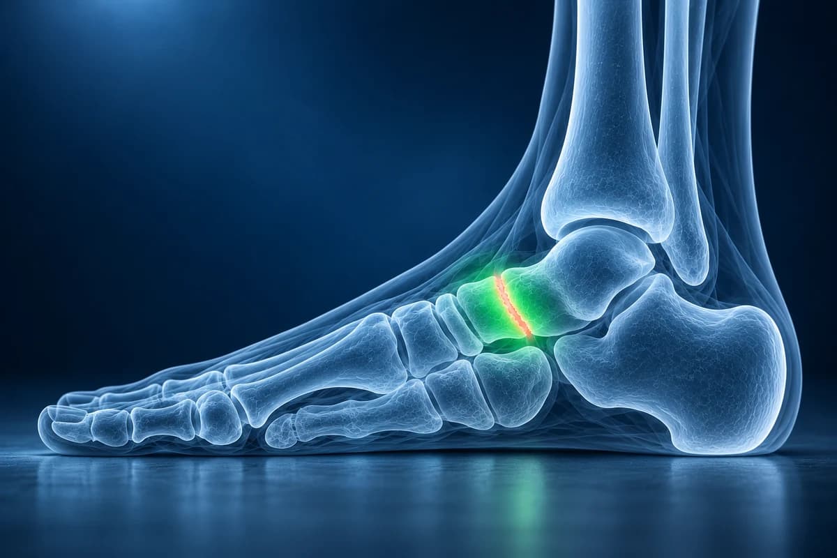
What matters more than the label is whether the arch is flexible or rigid, whether it is changing, and whether it hurts. This page explains the measurements a radiologist or foot surgeon writes about a flat foot, the patterns that deserve a closer look, and when a scan beyond the standing X-ray is useful. It is educational guidance, not a diagnosis.
Is a flat foot normal or a problem?
Most flat feet are a normal variation. Babies and toddlers have flat-looking feet, and the American Academy of Orthopaedic Surgeons notes that children with flexible flatfoot often do not develop an arch until 5 years of age or older. Many adults keep a low but flexible arch all their lives without pain. The children’s foot-pain guide puts concerns about a low arch in context.
A simple check separates the two main types. In a flexible flat foot the arch reappears when you sit, or when you rise onto your toes and the heel swings inward. In a rigid flat foot the arch stays flat and the heel stays tilted out, whatever you do. Rigid flat feet, flat feet that hurt, and an arch that collapses on one side in adult life are the ones that need assessment.
- A stiff, painful flat foot in an older child or teenager, often after sport or a minor sprain, can be caused by a tarsal coalition, a bridge between two hindfoot bones.
- A tender bump on the inner midfoot in a flexible flat foot may be an accessory navicular, an extra bone where the arch-supporting tendon attaches.
- A flat foot that develops in adulthood on one side, with pain and swelling behind the inner ankle, is most often caused by a failing posterior tibial tendon. Foot and ankle surgeons now call this progressive collapsing foot deformity and treat it as an ankle tendon problem first.
- A flat foot that appears after a twisting midfoot injury can signal a missed Lisfranc injury; a flat, swollen, warm foot in someone with diabetes and numb feet can signal Charcot foot.
When does a flat foot need urgent attention?
- You have diabetes or nerve damage and the foot becomes red, hot, swollen or changes shape over days or weeks. See a clinician the same day; an early Charcot foot needs offloading before the arch collapses further.
- You cannot bear weight on the midfoot after an injury, or there is bruising on the sole of the foot.
- The arch has visibly dropped on one side over a few months, with pain or swelling on the inside of the ankle.
- A child has a stiff, painful flat foot, limps, or has pain that wakes them at night.
What does “pes planus” mean on a foot X-ray report?
Flat feet are judged on weight-bearing X-rays, taken while you stand, because the arch can look normal when the foot is resting. These are the phrases you are most likely to see:
- “Meary's angle (lateral talar-first metatarsal angle) of 12°, apex plantar”: on the side view the axis of the talus points below the axis of the first metatarsal, so the line bends downward at the arch. Commonly used reference values read 0° to about 4° as normal, 4° to 15° as mild, 15° to 30° as moderate and more than 30° as severe pes planus.
- “Decreased calcaneal pitch”: the heel bone sits flatter than usual against the floor. A pitch of roughly 20° to 30° is typical, and values below about 18° to 20° fit a low arch.
- “Talonavicular uncoverage” or “increased talonavicular coverage angle”: on the top-down view the navicular has slid outward off the head of the talus, so part of the talar head is uncovered. It measures how much the forefoot has swung outward, called forefoot abduction.
- “Sag at the talonavicular or naviculocuneiform joint”: names the exact level where the arch has given way, which helps the surgeon plan if an operation is ever considered.
- “Hindfoot valgus” or “sinus tarsi impingement”: the heel leans outward, and in advanced cases the heel bone pinches against the tip of the fibula or the talus jams into the small tunnel on the outer side of the hindfoot, causing pain on the outside of the foot.
Angles shift by a few degrees between readers and between standing positions, so small changes are often within measurement error. How each line is drawn is shown in the foot X-ray angles guide, and the wider report is covered in how to read a foot X-ray. You can measure your own standing views in the free X-ray viewer. For pes planus wording beyond angle measurements, use the foot X-ray report glossary.
Do you need an MRI or CT for a flat foot?
A painless, flexible flat foot usually needs no imaging at all. When imaging is needed, standing X-rays of the foot (and often the ankle) come first, in line with the American College of Radiology criteria for chronic foot pain. They show the alignment, arthritis and bony causes such as a coalition or an extra navicular bone.
MRI is added when the cause may be a soft-tissue failure: the posterior tibial tendon, the spring ligament under the talar head, or bone bruising from impingement on the outer side. It also detects fibrous or cartilage coalitions that X-rays miss. CT shows bony coalitions and arthritis in fine detail, and weight-bearing CT, where available, shows the collapsed arch in three dimensions while you stand. For a side-by-side comparison, see foot MRI vs X-ray vs ultrasound; MRI files can be reviewed in the free MRI viewer.
What We Look For on Flat Foot Imaging
- Meary's angle and calcaneal pitch on the standing lateral view
- Talonavicular coverage and forefoot abduction on the standing top-down view
- The level of arch sag: talonavicular, naviculocuneiform or tarsometatarsal joints
- Bony causes such as a tarsal coalition or a type II accessory navicular
- Joint space narrowing and spurs that signal midfoot or hindfoot arthritis
- On MRI: posterior tibial tendon tearing, spring ligament laxity and impingement bone edema
How is an adult collapsing flat foot classified?
In 2020 a consensus group of foot and ankle surgeons replaced the older term adult acquired flatfoot deformity with progressive collapsing foot deformity. Their system first asks whether each part of the deformity is flexible or rigid, then names where the collapse is: the heel tilting outward, the forefoot swinging outward at the talonavicular joint, the inner column of the midfoot giving way, the talus slipping off the bones beneath it, or the ankle itself tilting. The combination guides whether bracing, physical therapy or surgery is offered.
A low arch can also change how load passes through the rest of the foot. Some people with flat feet develop heel pain from strain on the plantar fascia or a bunion at the big toe, but many never do.
Frequently Asked Questions
What Meary's angle counts as a flat foot?
On a standing side-view X-ray, the talus and first metatarsal normally line up within about 4°. An angle that bends downward by more than about 4° is read as pes planus; commonly used bands call 4° to 15° mild, 15° to 30° moderate and above 30° severe. The number supports, but does not replace, the clinical examination.
Should a flat foot X-ray be taken standing?
Yes. The arch flattens under body weight, so X-rays taken lying down can make a flat foot look normal. Ask whether your images were weight-bearing before comparing angles between studies.
Do flat feet in children need treatment?
Most flexible, painless flat feet in children do not, because the arch often forms gradually through childhood. A child with pain, stiffness, a limp or one foot clearly different from the other should be examined, and an X-ray may then look for a coalition or an accessory navicular.
Can flat feet get worse in adults?
A long-standing flexible flat foot often stays the same for years. An arch that drops on one side in adult life, especially with pain on the inside of the ankle, is different: it can progress and becomes harder to correct once rigid, so it is worth having assessed early.
Can AI measure my flat foot X-ray?
Weight-bearing X-rays show arch alignment, while examination checks flexibility and tendon function. AI cannot provide validated measurements or choose treatment.
Sources
- Moore J, Tafti D. Pes Planus. In: StatPearls. Treasure Island (FL): StatPearls Publishing.
- Myerson MS, Thordarson DB, Johnson JE, et al. Classification and Nomenclature: Progressive Collapsing Foot Deformity. Foot Ankle Int. 2020;41(10):1271-1276.
- Expert Panel on Musculoskeletal Imaging; Said N, Fox MG, Nacey NC, et al. ACR Appropriateness Criteria Chronic Foot Pain: Update 2025. J Am Coll Radiol. 2026;23(4):681-699.
- American Academy of Orthopaedic Surgeons. Flexible Flatfoot in Children. OrthoInfo.
Related Articles
Why Lisfranc midfoot injuries are missed on first X-rays, the alignment lines and fleck sign to check, and when standing X-rays, CT or MRI are needed.
How long plantar fasciitis lasts, what changes month by month, why morning pain is the last to go, and when imaging or a different diagnosis should be considered.
What the numbers on your foot X-ray mean: hallux valgus angle, intermetatarsal angle, Meary's angle, calcaneal pitch and Böhler's angle, with normal ranges and how each is measured.
Understand your imaging before your next appointment
MRI, X-ray or CT from a CD, folder or ZIP. A PDF report or photos of the films work too.
Free to open and preview, no account needed. An optional AI report from $10 explains your images in plain language, with questions for your doctor.
Your raw files stay on this device. Only rendered images and minimal context are sent when you request an AI report.
Medical Disclaimer: This page is for informational and educational purposes only. It does not constitute medical advice, diagnosis, or treatment. AI-generated analysis may contain errors. Always consult a qualified healthcare professional for medical decisions. Full Disclaimer