
Lisfranc Injury Missed on X-Ray: Signs, Standing Views and MRI
Why Lisfranc midfoot injuries are missed on first X-rays, the alignment lines and fleck sign to check, and when standing X-rays, CT or MRI are needed.
Key Takeaways
- As many as 20% of Lisfranc injuries have been reported missed on initial non-weight-bearing X-rays
- Ligament-only injuries may only show a gap when the foot is X-rayed standing
- The second metatarsal base should line up with the middle cuneiform; a widened gap compared with the other foot is suspicious
- A fleck sign is a small avulsed bone chip that points to a significant ligament injury
- CT details fractures for surgery; MRI shows the ligament when X-rays are normal
- Bruising on the sole after a midfoot twist should prompt a check for a Lisfranc injury
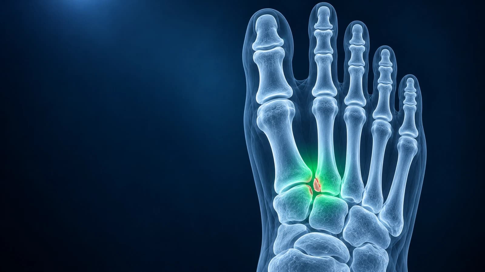
Lisfranc injuries are among the most commonly missed foot injuries: a classic review estimated that as many as 20% are missed on the first non-weight-bearing X-rays. A ligament-only injury can leave the bones lined up while you lie on the table, and only separate when the foot is loaded. Standing X-rays, a comparison with the other foot, CT or MRI uncover what the first films did not.
The injury is easily mistaken for a midfoot sprain, and the AAOS warns it should not simply be walked off. Bruising on the sole of the foot after a twist is a strong warning sign. This guide explains why the first X-ray can look normal, the alignment checks and report phrases that matter, and when further imaging is needed. It is educational and not a diagnosis; if this sounds like your injury, see a clinician.
Why can a Lisfranc injury look normal on X-ray?
The Lisfranc joint complex is the row of joints where the five metatarsals meet the midfoot bones. Its keystone is the base of the second metatarsal, which sits recessed between the cuneiforms like a key in a lock. Unusually, there is no strong ligament tying the bases of the first and second metatarsals together. Instead, the Lisfranc ligament runs diagonally from the medial cuneiform to the base of the second metatarsal and holds the arch together.
When that ligament is torn but no bone is broken, the joints can sit in their normal position while the foot is unloaded. A routine foot X-ray is often taken lying down or sitting, so the gap that would open under body weight never appears. Swelling and pain also make it hard to stand for proper views, the bones overlap heavily in the midfoot, and a subtle shift of one or two millimeters is easy to overlook in a busy emergency department.
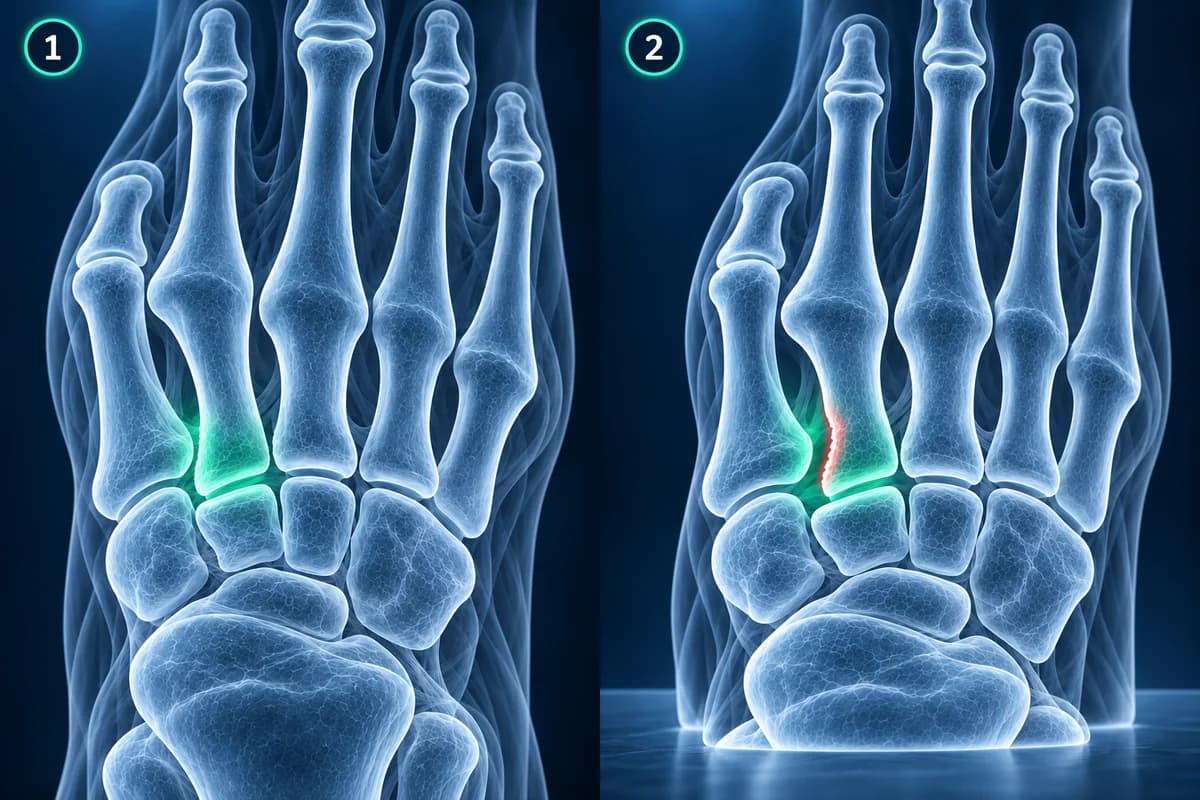
What should be checked on a foot X-ray for a Lisfranc injury?
Radiologists look at a few alignment lines that should be continuous on every normal foot. On the front-to-back view, the inner edge of the second metatarsal base should line up with the inner edge of the middle cuneiform. On the oblique view, the inner edge of the fourth metatarsal base should line up with the inner edge of the cuboid. On the side view, the tops of the metatarsal bases should not step up above the midfoot bones.
They also measure the space between the base of the first and second metatarsals and between the medial cuneiform and the second metatarsal. A gap larger than on the uninjured foot, often around 2 mm or more, is suspicious, which is why a standing comparison view of both feet is so useful. The guide to how to read a foot X-ray and the explainer on foot X-ray angles show these views in context, and you can open your own films in the free X-ray viewer.
What does "fleck sign" mean on a foot X-ray?
"Fleck sign" describes a tiny chip of bone in the space between the bases of the first and second metatarsals. It is usually a piece pulled off by the Lisfranc ligament as it tore, so a small fleck points to a significant ligament injury and possible instability, even when everything else looks aligned. It deserves the same attention as an obvious fracture.
Other phrases you may meet: "widening of the first intermetatarsal space at the base" or "diastasis between the medial cuneiform and second metatarsal base" means the bones have separated more than expected. "Malalignment at the second tarsometatarsal joint" means the inner edges no longer line up. "Dorsal step-off" means a metatarsal base has shifted upward on the side view. On MRI, "disruption" or "high-grade sprain of the Lisfranc ligament" describes a torn ligament, and "bone marrow edema in the medial cuneiform and second metatarsal base" describes bone bruising at its attachments.
When are standing X-rays, CT or MRI needed?
The American College of Radiology criteria for acute foot trauma recommend three-view radiographs for a suspected Lisfranc injury, with weight bearing on at least the front-to-back view. If those films are normal but the injury is still suspected, MRI or CT may be appropriate. When pain prevents standing, a short period of protection followed by repeat standing views is sometimes used instead.
CT shows small fractures, fragments and joint surfaces in fine detail and is commonly used to plan surgery. MRI shows the ligament itself, so it is the test that can separate a stretched ligament from a complete tear when the X-rays and CT show no fracture. Neither replaces the standing X-ray as a test of stability; they answer different questions. Our comparison of foot MRI vs X-ray vs ultrasound and the guide to reading a foot MRI go further.
Which symptoms should make you suspect a missed Lisfranc injury?
The typical story is a twist or fall onto a foot pointed downward, such as being landed on in a tackle, missing a step, or catching the foot in a stirrup or pedal; high-energy car and work accidents cause the more severe forms. The pain and swelling sit on the top of the midfoot rather than at the ankle. Standing on tiptoe is very painful or impossible, and bruising may appear on the sole a day or two later.
If you were told it was a sprain but you still cannot push off, the midfoot stays swollen after a couple of weeks, or walking feels unstable, ask whether standing X-rays of both feet or further imaging have been done. That question is reasonable even when the first X-ray report was normal.
Why does a missed Lisfranc injury matter?
Treatment depends on stability. According to the AAOS, injuries without fractures, dislocation or a complete ligament tear are usually treated in a non-weight-bearing cast or boot for 6 to 8 weeks, then a gradual return to walking. Displaced injuries are usually treated with surgery, either fixation with screws or plates or a fusion of the affected joints.
When an unstable injury is treated as a sprain, the midfoot can slowly drift out of position. The result can be long-term pain, arthritis of the midfoot joints and a gradual collapse of the arch that resembles an acquired flat foot. Recognizing the injury early keeps more options open. The Lisfranc injury condition page covers the full range of injuries and treatment. Pain at the base of the second metatarsal without a twist can instead be a metatarsal stress fracture, which is also easy to miss on early films.
When is a midfoot injury an emergency?
Go to emergency care straight away if you cannot bear any weight, if the foot is visibly deformed, if the toes turn pale, blue, cold or numb, or if the swelling is tense and the pain is severe and getting worse despite rest and elevation. High-energy midfoot injuries can cause dangerous pressure inside the foot that needs urgent treatment. An open wound over the injury also needs same-day care.
Frequently Asked Questions
Can a Lisfranc injury be missed on X-ray?
Yes. Subtle and ligament-only injuries are frequently missed, especially when the X-rays are taken without weight bearing. A widely cited review reported that as many as 20% are missed on initial films. Standing views of both feet, CT or MRI are used when the suspicion remains.
Will a weight-bearing X-ray make a Lisfranc injury worse?
The AAOS notes that standing X-rays will not worsen the injury. Weight bearing is what reveals an unstable ligament injury, so it is a key part of the workup. If pain makes standing impossible, your clinician may protect the foot first and repeat the views later.
Is MRI or CT better for a suspected Lisfranc injury?
They answer different questions. CT is better for small fractures and for planning surgery. MRI is better for seeing the Lisfranc ligament itself and bone bruising when no fracture is visible. Many people need only standing X-rays; the extra tests are added when the answer is still unclear.
How do I know if my midfoot sprain is actually a Lisfranc injury?
You cannot tell reliably on your own. Warning signs include bruising on the sole, inability to stand on tiptoe, swelling over the top of the midfoot, and pain that does not ease over the first couple of weeks. If any of these apply, ask your clinician whether standing X-rays or further imaging are needed.
What if my Lisfranc injury was diagnosed late?
A late diagnosis does not remove treatment options, but the choices can change. Depending on alignment and symptoms, options range from protected weight bearing and supportive inserts to fixation or fusion. A foot and ankle specialist can review your imaging and explain what applies to you.
Sources
- Burroughs KE, Reimer CD, Fields KB. Lisfranc Injury of the Foot: A Commonly Missed Diagnosis. Am Fam Physician. 1998;58(1):118-124.
- Expert Panel on Musculoskeletal Imaging; Gorbachova T, Chang EY, Ha AS, et al. ACR Appropriateness Criteria Acute Trauma to the Foot. J Am Coll Radiol. 2020;17(5S):S2-S11.
- American Academy of Orthopaedic Surgeons. Lisfranc (Midfoot) Injury. OrthoInfo.
- Stern JM, Bergman R, Singh S. Lisfranc Dislocation. In: StatPearls [Internet]. Treasure Island (FL): StatPearls Publishing.
Related Articles
A plain-language guide to your foot X-ray: the three standard views, how to trace alignment from heel to toes, and what fractures, fleck signs, accessory bones and arthritis look like.
What the numbers on your foot X-ray mean: hallux valgus angle, intermetatarsal angle, Meary's angle, calcaneal pitch and Böhler's angle, with normal ranges and how each is measured.
X-ray, MRI, ultrasound or CT for foot pain? What each test shows and misses, which one usually comes first, and how the choice changes for stress fractures, neuromas, plantar fasciitis, Lisfranc injuries and the diabetic foot.
Related Conditions
Understand your imaging before your next appointment
MRI, X-ray or CT from a CD, folder or ZIP. A PDF report or photos of the films work too.
Free to open and preview, no account needed. An optional AI report from $10 explains your images in plain language, with questions for your doctor.
Your raw files stay on this device. Only rendered images and minimal context are sent when you request an AI report.
Medical Disclaimer: This page is for informational and educational purposes only. It does not constitute medical advice, diagnosis, or treatment. AI-generated analysis may contain errors. Always consult a qualified healthcare professional for medical decisions. Full Disclaimer