
Plantar Fasciitis: When Imaging Helps and What Your Report Means
Most people with plantar fasciitis do not need a scan. The diagnosis is made from your story and an examination: sharp pain under the inner heel with the first morning steps and a tender point where the fascia meets the heel bone. Imaging is added when pain lasts despite months of good care or another cause is suspected. On ultrasound, a fascia thicker than about 4 mm at the heel supports the diagnosis.
Common Symptoms
- Sharp, stabbing pain under the inner side of the heel with the first steps out of bed in the morning
- Start-up pain that returns after sitting for a while, then eases as you keep walking
- A tender spot on the bottom of the heel where the plantar fascia attaches to the heel bone
- Pain that builds again after long periods of standing, especially on hard floors
- Heel pain that worsens when the big toe is bent upward, stretching the fascia along the arch
- Tight calf muscles and difficulty pulling the foot and ankle upward toward the shin
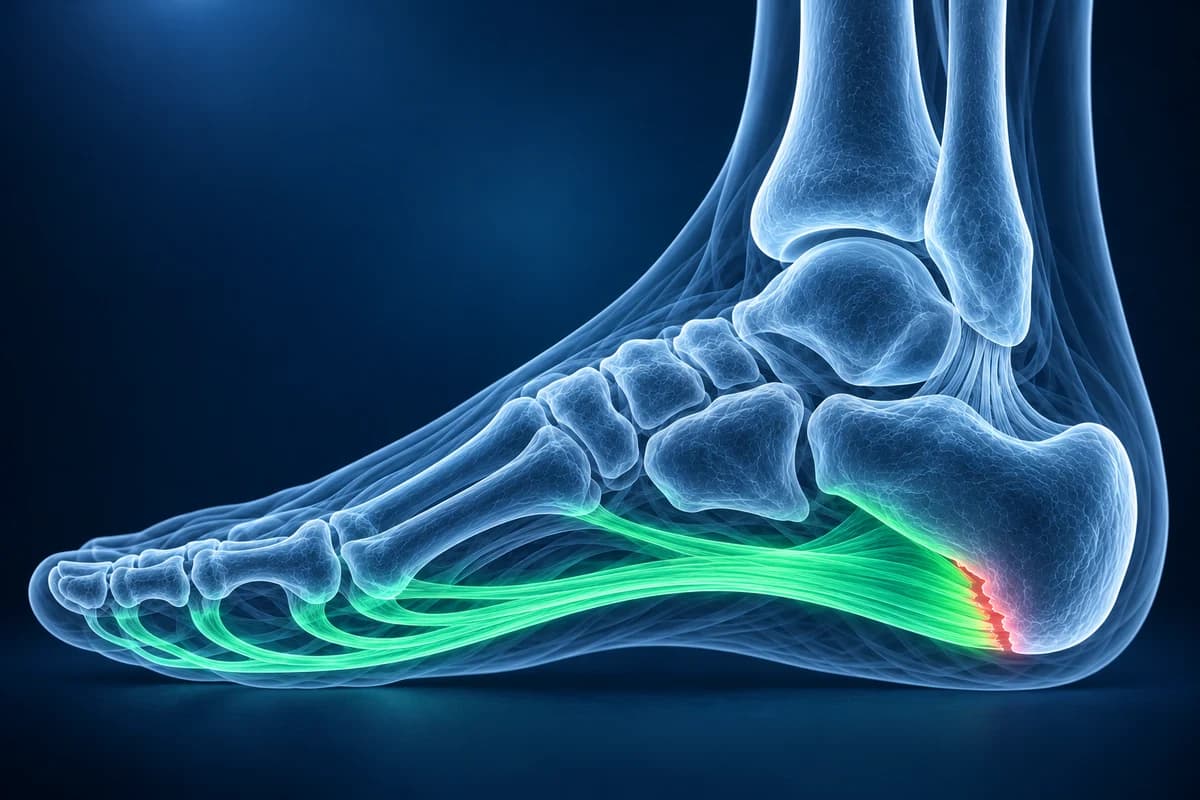
The plantar fascia is a thick band of connective tissue that holds up the arch like a bowstring. Repeated strain where it anchors to the heel causes small-scale degeneration rather than true inflammation, which is why many specialists now prefer the word fasciopathy. This page explains when an X-ray, ultrasound or MRI is genuinely useful, the phrases those reports use, and the heel pain patterns that point to something else. It is educational guidance, not a diagnosis.
Do you need an X-ray or MRI for plantar fasciitis?
Usually not at the first visit. Physical therapy clinical practice guidelines on heel pain treat plantar fasciitis as a clinical diagnosis, and the standard first steps (calf and fascia stretching, supportive shoes, temporary reduction of impact, load management and sometimes a night splint) do not depend on what a scan shows. The American Academy of Orthopaedic Surgeons notes that more than 90% of people improve within 10 months of starting these simple treatments.
Imaging becomes reasonable when heel pain has not settled after several months of consistent treatment, when the story does not fit (pain after a fall, night pain, swelling, numbness), or before a procedure such as an injection, shockwave therapy or surgery. For chronic foot pain, the American College of Radiology Appropriateness Criteria place plain X-rays first and keep ultrasound and MRI for the questions X-rays cannot answer. The full decision is walked through in do you need an MRI for plantar fasciitis, and what to expect week by week is in the plantar fasciitis recovery timeline.
When is heel pain not plantar fasciitis? Red flags
Seek care promptly, and the same day for the first three, if any of these apply. They are reasons to be examined, not reasons to wait for heel stretches to work.
- You cannot put weight on the heel after a fall, a jump from height or a car accident; this needs an X-ray to rule out a calcaneal fracture.
- You have diabetes and the foot is suddenly hot, red or swollen, or there is a wound, fever or pus. These can mean infection such as diabetic foot osteomyelitis or an active Charcot foot.
- Fever, chills or a heel that is warm and swollen all over rather than tender in one spot.
- Pain that started after a jump in running mileage and hurts when the sides of the heel are squeezed, which suggests a calcaneal stress fracture rather than fascia strain.
- Burning, tingling or numbness in the sole can suggest a nerve problem, such as tarsal tunnel syndrome behind the inner ankle; ask your clinician.
- Pain at rest or at night, pain in both heels together with morning back stiffness, or swollen toes, which can be signs of an inflammatory arthritis.
Pain at the back of the heel, where the Achilles tendon inserts, is a different problem from plantar fasciitis and is assessed as an ankle and tendon condition.
What does “thickened plantar fascia” mean on an ultrasound or MRI report?
These are the phrases heel imaging reports print most often, with what each one means in plain words. Our MRI report phrase guide provides context for plantar fascia wording.
- “Thickening of the proximal plantar fascia, measuring 6 mm”: the band is swollen where it leaves the heel. A healthy fascia is usually under 4 mm thick there, and a meta-analysis found that thickness above 4.0 mm on ultrasound strongly favors plantar fasciitis.
- “Hypoechoic” or “loss of the normal fibrillar pattern”: on ultrasound the fascia looks darker and less striped than normal, a sign of degenerated, waterlogged collagen.
- “Perifascial edema” or “increased T2 signal at the calcaneal origin”: fluid around or within the fascia on MRI, the soft-tissue footprint of active irritation.
- “Bone marrow edema at the medial calcaneal tuberosity”: a stress reaction in the heel bone right where the fascia pulls. It often accompanies stubborn plantar fasciitis but is also checked carefully for an early stress fracture.
- “Plantar calcaneal enthesophyte” or “plantar heel spur”: a beak of bone on the underside of the heel, discussed in its own section below.
- “Partial-thickness tear of the plantar fascia”: some fibers have torn, sometimes after a sudden pop or after a steroid injection. A rupture can change how long you protect the foot.
A firm lump in the middle of the arch, reported as a “nodule within the plantar fascia”, is usually a plantar fibroma. It is a separate, benign condition and is treated differently from heel-origin fasciitis. Consult the heel-pain guide for symptoms outside the usual plantar fascia pattern.
Does a heel spur on an X-ray mean the spur is causing your pain?
Not necessarily. A plantar heel spur is common and many people who have one never feel heel pain. In the meta-analysis by McMillan and colleagues, spurs were clearly more frequent in people with chronic heel pain than in people without it, so they are linked, but a spur on its own does not prove that it is the source of pain. The spur often sits just above the fascia, in the origin of the small toe-flexor muscles, and grows slowly over years of traction.
In practice, treatment targets the strained fascia and calf, not the spur, and the spur usually stays on later X-rays even after the pain has gone. Surgery to remove a spur is rarely needed. If your report mentions a spur and nothing else, that fits ordinary plantar fasciitis; the X-ray was mainly useful because it ruled out a fracture, a bone lesion or arthritis.
What can each test show for heel pain?
X-ray is quick and cheap. It shows bone: spurs, fractures, arthritis, bone cysts and, sometimes, the healing line of a stress fracture. It cannot show the fascia itself, and a fresh calcaneal stress fracture often looks normal in the first weeks; the guide to stress fractures that do not show on X-ray explains why. If you already have the images, you can open them in the free X-ray viewer.
Ultrasound measures fascia thickness, shows darker degenerated fibers, detects small tears and can guide a needle if an injection is planned. It is fast and radiation-free, but the result depends on the operator.
MRI is the most complete test. It shows the fascia, fluid around it, bone marrow edema in the heel, a stress fracture line, nerve entrapment and rarer causes such as a tumor or infection. It is mainly worth doing when the diagnosis is uncertain or before surgery. An MRI you received on a disc or as DICOM files can be opened in the free MRI viewer, and the foot MRI vs X-ray vs ultrasound comparison covers costs and trade-offs.
What We Look For on Heel Imaging
- Plantar fascia thickness at the heel origin, compared with the 4 mm reference point
- Fluid around the fascia or increased signal within it on fluid-sensitive MRI sequences
- Partial-thickness or full-thickness fascia tears and their distance from the heel
- Bone marrow edema at the medial calcaneal tuberosity and any stress fracture line in the heel bone
- Plantar heel spur size, and whether it sits within or above the fascia origin
- Alternative causes such as heel pad atrophy, a bone cyst, nerve compression or a plantar fibroma
Does foot shape make plantar fasciitis more likely?
Both a low arch and a high arch can increase strain on the fascia, as can tight calves, a sudden rise in standing or running, body weight and age between about 40 and 60. If your X-ray report also describes a collapsed arch, the flat foot page explains the angles used to measure it. Foot shape is one factor among several, and arch supports help some people more than others.
Frequently Asked Questions
Can an X-ray show plantar fasciitis?
Not directly. X-rays show bone, not the fascia, so a normal X-ray is common with plantar fasciitis. An X-ray may show a heel spur or blurring of the fat under the heel, and it helps rule out a fracture, a bone cyst or arthritis. Ultrasound or MRI is needed to see the fascia itself.
How thick is the plantar fascia in plantar fasciitis?
A healthy plantar fascia is usually less than 4 mm thick where it leaves the heel. A systematic review of imaging studies found that thickness above 4.0 mm on ultrasound strongly favors plantar fasciitis, and painful fascia were on average about 2 mm thicker than in people without heel pain. Thickness is supporting evidence; your clinician still makes the diagnosis from the examination.
When should I get an MRI for heel pain?
Typically when pain continues after several months of consistent treatment, when a stress fracture, tear, nerve problem or other cause is suspected, or before an operation. An MRI early in typical plantar fasciitis rarely changes the treatment plan. Your clinician decides whether the scan is worth it in your case.
Should a heel spur be removed?
Rarely. Spurs are common in people without any heel pain and usually remain after the pain has settled. Treatment focuses on the fascia and the calf. Surgery for stubborn heel pain is considered only after many months of non-surgical care, and spur removal is not its main goal.
Can AI read my heel MRI or ultrasound report?
Fascia thickening and surrounding edema may support plantar fasciitis, but heel pain has other causes and needs examination. AI cannot confirm the diagnosis.
Sources
- McMillan AM, Landorf KB, Barrett JT, Menz HB, Bird AR. Diagnostic imaging for chronic plantar heel pain: a systematic review and meta-analysis. J Foot Ankle Res. 2009;2:32.
- Koc TA Jr, Bise CG, Neville C, Carreira D, Martin RL, McDonough CM. Heel Pain - Plantar Fasciitis: Revision 2023. J Orthop Sports Phys Ther. 2023;53(12):CPG1-CPG39.
- Expert Panel on Musculoskeletal Imaging; Said N, Fox MG, Nacey NC, et al. ACR Appropriateness Criteria Chronic Foot Pain: Update 2025. J Am Coll Radiol. 2026;23(4):681-699.
- American Academy of Orthopaedic Surgeons. Plantar Fasciitis and Bone Spurs. OrthoInfo.
Related Articles
How long plantar fasciitis lasts, what changes month by month, why morning pain is the last to go, and when imaging or a different diagnosis should be considered.
Heel pain mapped to its causes: plantar fasciitis, heel stress fracture, Baxter's nerve, fat pad atrophy and Sever's disease, and which test shows each one.
Most plantar fasciitis needs no MRI. Learn when an X-ray, ultrasound or MRI is worth it, what each shows, and what heel pain report phrases mean.
Understand your imaging before your next appointment
MRI, X-ray or CT from a CD, folder or ZIP. A PDF report or photos of the films work too.
Free to open and preview, no account needed. An optional AI report from $10 explains your images in plain language, with questions for your doctor.
Your raw files stay on this device. Only rendered images and minimal context are sent when you request an AI report.
Medical Disclaimer: This page is for informational and educational purposes only. It does not constitute medical advice, diagnosis, or treatment. AI-generated analysis may contain errors. Always consult a qualified healthcare professional for medical decisions. Full Disclaimer