
Do You Need an MRI for Plantar Fasciitis?
Most plantar fasciitis needs no MRI. Learn when an X-ray, ultrasound or MRI is worth it, what each shows, and what heel pain report phrases mean.
Key Takeaways
- Typical plantar fasciitis is diagnosed by history and examination, without an MRI
- More than 90% of people improve within 10 months of simple treatment, according to the AAOS
- X-rays rule out other causes; a heel spur on the report is common and does not cause the pain
- Ultrasound measures fascia thickness, with about 4 mm the usual cut-off for thickening
- MRI is for atypical pain, failed treatment, suspected stress fracture, tear or nerve problem, or surgical planning
- Inability to bear weight, fever, or a hot swollen diabetic foot needs urgent care
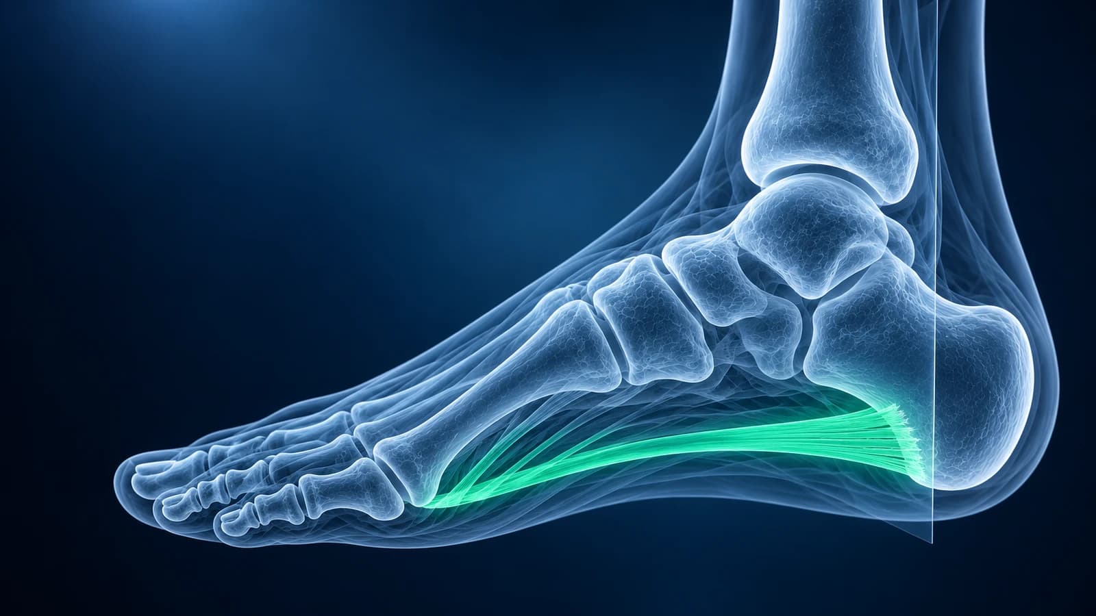
Usually, no. Plantar fasciitis is diagnosed from your story and an examination: sharp pain under the inner heel with the first steps in the morning or after sitting, and tenderness where the fascia meets the heel bone.
The American Academy of Orthopaedic Surgeons notes that MRI and ultrasound are not routinely used, and that more than 90% of people improve within 10 months of simple treatment. Imaging becomes useful when the pain does not fit that pattern, does not respond to good care, or another cause is suspected.
That answer surprises people who have been in pain for weeks and want to see what is wrong. The reason is simple: for typical heel pain, a scan rarely changes the plan. Stretching, load management, supportive footwear, and time are the treatment whether the fascia measures slightly thick or very thick. Below is how clinicians decide when a test is worth it, what each test can show, and how to read the phrases that appear in heel imaging reports. It is general education and not a diagnosis for your own foot.
Why is plantar fasciitis usually diagnosed without imaging?
The symptoms of plantar fasciitis are unusually consistent. Pain is worst with the first steps after rest, eases as the foot warms up, and returns after long periods on the feet. The tender spot sits at the front and inner side of the heel, not in the middle of the heel pad and not along the back of the heel. Pulling the toes up toward the shin often reproduces it, because that tightens the fascia like a bowstring.
When those features line up in an adult with a recent change in activity, weight, footwear or standing time, the diagnosis is clinical, and the first-line treatment is the same with or without a scan. Ordering an MRI at this stage adds cost and waiting time, and it can find incidental changes that worry people without changing what they should do.
When is an MRI or other scan worth it for heel pain?
Imaging is reasonable when the story has a twist. Common triggers for a scan include:
- No real improvement after several months of consistent treatment, or before an injection, shockwave therapy or surgery is planned
- Pain that started with a fall, a jump or a sudden pop, or that is felt when the heel is squeezed from both sides, which raises the question of a calcaneal stress fracture
- Burning, tingling or numbness in the sole, which points toward a trapped nerve rather than the fascia
- Pain at night or at rest, swelling, a lump, or heel pain on both sides with other joint symptoms
- Sudden worsening with bruising in the arch, especially after a steroid injection
- Heel pain in a child or teenager, where growth-plate problems are more likely than fasciitis
In these cases the American College of Radiology chronic foot pain criteria favor starting with X-rays, then adding ultrasound or MRI depending on the question. MRI is the broadest test: it shows the fascia, the heel bone marrow, the fat pad, nearby tendons and nerves in one study.
What can an X-ray show in plantar fasciitis?
An X-ray cannot see the fascia itself, which is soft tissue. Its job is to rule out other causes: a stress fracture that has started to heal and shows as a dense band across the heel bone, arthritis, a bone cyst or tumor, or the signs of inflammatory arthritis at tendon attachments. It is also the test that shows a heel spur.
A heel spur on the report does not mean the spur is causing your pain. Spurs are common in people with no heel pain at all, and the AAOS states plainly that heel spurs do not cause plantar fasciitis pain. Treatment targets the fascia, not the spur. If you want to look at your own films, our guide on how to read a foot X-ray explains the lateral heel view, and you can open the images in the free X-ray viewer.
Is ultrasound or MRI better for plantar fasciitis?
Ultrasound is the practical choice when the question is about the fascia itself. It measures the thickness of the fascia where it attaches to the heel and compares it with the other foot; a thickness above about 4 mm is the commonly used cut-off for a thickened fascia. It also shows a darker, swollen texture, fluid around the fascia, and some tears. It is quick, has no radiation, is often cheaper, and can guide an injection to the right spot.
MRI is the choice when the question is wider. It can show thickening and abnormal signal in the fascia, but its real strength is seeing everything around it: swelling inside the heel bone from a stress fracture, bruising of the bone where the fascia pulls, a partial or complete tear, a damaged fat pad, or changes in the nerves and tendons nearby. Our comparison of foot MRI vs X-ray vs ultrasound covers the trade-offs for other foot problems too.
What do plantar fasciitis report phrases mean?
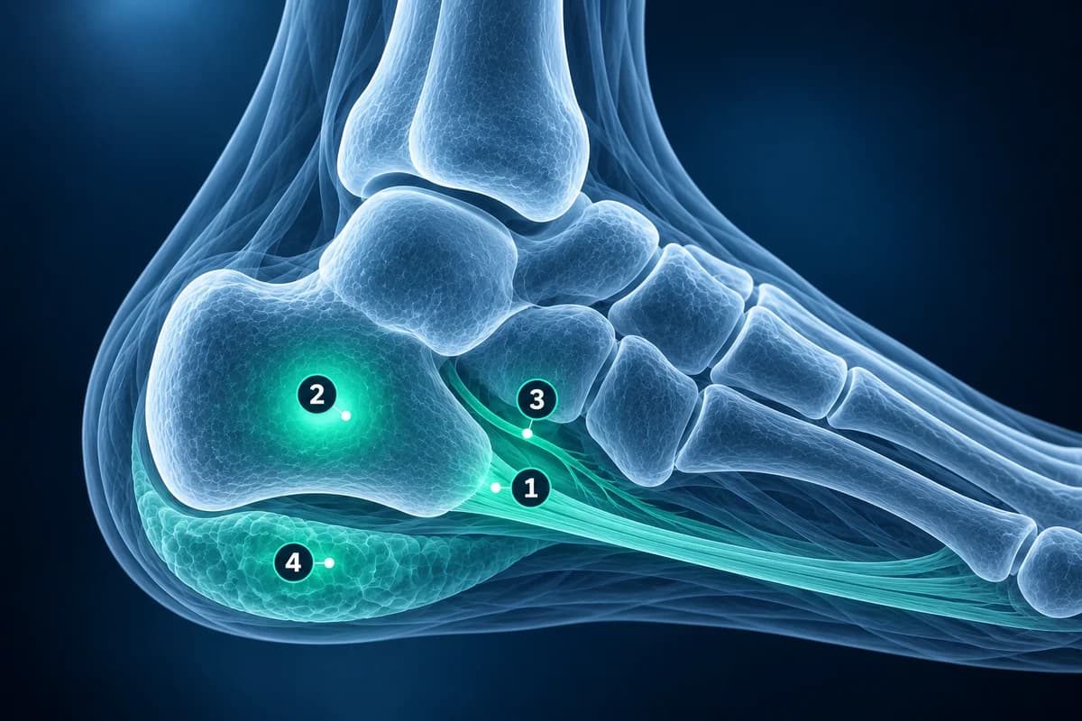
"Thickening of the proximal plantar fascia" means the fascia is thicker than expected where it attaches to the heel, the hallmark imaging sign of plantar fasciitis. "Intermediate or increased T2 signal within the fascia" means the fibers hold more water than healthy fascia, a sign of irritation or degeneration.
"Perifascial edema" is fluid in the soft tissue around the fascia, which fits an active, painful phase. "Bone marrow edema at the medial calcaneal tubercle" describes stress in the bone right where the fascia pulls, and it can be part of plantar fasciitis or point toward a stress injury of the heel bone.
"Plantar calcaneal enthesophyte" is the formal name for a heel spur. "Partial-thickness tear" or "rupture of the plantar fascia" means some or all fibers have torn, which changes the treatment and is worth discussing with your clinician. "Fat pad atrophy" or "fat pad edema" points to the cushion under the heel, a different cause of heel pain that needs a different approach.
What else can feel like plantar fasciitis?
Several problems share the heel. A calcaneal stress fracture tends to hurt more as the day goes on and when the heel is squeezed, rather than mainly with the first steps; early X-rays can be normal, which is the theme of our article on a stress fracture that is not on the X-ray. A high-energy fall onto the heel can cause a calcaneal fracture, which needs prompt X-rays and often CT.
Nerve entrapment around the inner ankle and heel causes burning and numbness rather than first-step pain. Pain at the back of the heel usually comes from the Achilles tendon insertion, an ankle-region problem rather than the fascia. Foot shape matters too: a flat foot loads the fascia differently, and in teenagers a stiff flat foot can come from a tarsal coalition. Heel pain on both sides together with back, eye or other joint symptoms can reflect inflammatory arthritis.
When should heel pain be seen urgently?
Get prompt medical care if you cannot put weight on the heel after a fall or jump, if the heel is hot, red and swollen, if you have a fever, if you have diabetes and notice a wound, swelling or color change on the foot, or if numbness is spreading. These are not features of ordinary plantar fasciitis and should not wait for a scan appointment.
Already have a heel scan?
If you already have an ultrasound, X-ray or MRI of your heel, you can open it privately in the free MRI viewer or upload it for an educational explanation of what the images and report describe. For what to expect over the coming months, read our plantar fasciitis recovery timeline.
Frequently Asked Questions
Can an MRI confirm plantar fasciitis?
An MRI can show changes that fit plantar fasciitis, such as a thickened proximal fascia with increased signal and swelling around it. But the diagnosis is still made by matching those findings to your symptoms and examination. Thickening can be present in people with little pain, so the scan supports the diagnosis rather than making it on its own.
How long should I wait before asking for imaging for heel pain?
For typical plantar fasciitis without warning signs, most clinicians try several months of consistent treatment first. Imaging comes earlier if the pain followed an injury, includes numbness, is present at rest or at night, comes with swelling or fever, or affects a child. Your clinician can weigh those factors for your situation.
Does a heel spur on my X-ray mean I need surgery?
No. Heel spurs are common in people without pain, and the AAOS notes they do not cause plantar fasciitis pain. Surgery for plantar fasciitis is generally reserved for people who have not improved after about 12 months of consistent nonsurgical treatment, and it addresses the fascia rather than the spur.
Is ultrasound enough for plantar fasciitis?
Often, yes, when the question is only about the fascia. Ultrasound shows thickness, texture, fluid and many tears, and can guide an injection. If the pain may come from the heel bone, a nerve or another structure, MRI gives the broader view.
What does bone marrow edema in the heel mean on an MRI?
It means there is extra fluid inside the heel bone. A small area right where the fascia attaches can go along with plantar fasciitis. A larger area, or a line through the heel bone, raises the possibility of a stress reaction or stress fracture, which is managed differently, so it is worth reviewing with your clinician.
Sources
- American Academy of Orthopaedic Surgeons. Plantar Fasciitis and Bone Spurs. OrthoInfo.
- Plantar Fasciitis. In: StatPearls [Internet]. Treasure Island (FL): StatPearls Publishing.
- Expert Panel on Musculoskeletal Imaging; Said N, Fox MG, Nacey NC, et al. ACR Appropriateness Criteria Chronic Foot Pain: Update 2025. J Am Coll Radiol. 2026;23(4):681-699.
Related Articles
How long plantar fasciitis lasts, what changes month by month, why morning pain is the last to go, and when imaging or a different diagnosis should be considered.
What a foot MRI typically costs, why forefoot, midfoot and contrast scans are billed differently, how insurance applies, and how to lower your bill.
X-ray, MRI, ultrasound or CT for foot pain? What each test shows and misses, which one usually comes first, and how the choice changes for stress fractures, neuromas, plantar fasciitis, Lisfranc injuries and the diabetic foot.
Related Conditions
Understand your imaging before your next appointment
MRI, X-ray or CT from a CD, folder or ZIP. A PDF report or photos of the films work too.
Free to open and preview, no account needed. An optional AI report from $10 explains your images in plain language, with questions for your doctor.
Your raw files stay on this device. Only rendered images and minimal context are sent when you request an AI report.
Medical Disclaimer: This page is for informational and educational purposes only. It does not constitute medical advice, diagnosis, or treatment. AI-generated analysis may contain errors. Always consult a qualified healthcare professional for medical decisions. Full Disclaimer