
How to Read a Foot X-ray: Views, Fractures and Report Terms
A plain-language guide to your foot X-ray: the three standard views, how to trace alignment from heel to toes, and what fractures, fleck signs, accessory bones and arthritis look like.
Key Takeaways
- A foot series has three views; standing films are needed to judge bunions, arches and midfoot gaps
- Read in order: alignment from heel to toes, then each bone outline, then joints and soft tissues
- The second metatarsal should line up with the middle cuneiform; a step or fleck suggests a Lisfranc injury
- Accessory bones and bipartite sesamoids have smooth edges, fresh fractures have sharp ones
- A normal X-ray does not rule out a stress fracture, neuroma, plantar plate or plantar fascia problem

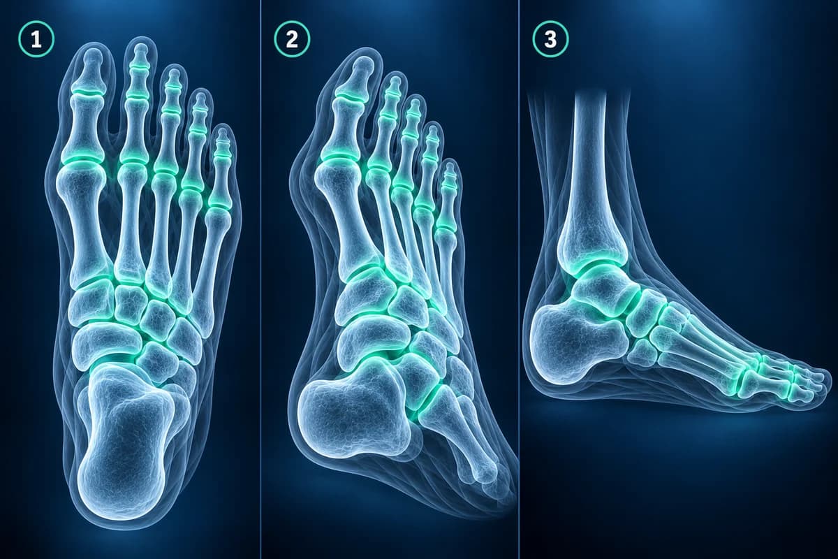
To read a foot X-ray, look at three things in order: whether the 26 bones of the foot line up, whether any bone outline is broken, and whether the joints are narrowed or worn. A standard foot series has three views (top-down, oblique and side), and a standing film is needed to judge alignment and deformity such as a bunion or flat foot.
This guide explains what each view is for, the order radiologists follow, and the phrases that appear in a typical foot X-ray report. It is educational and not a diagnosis: the person who examined your foot can match the image to the tender spot, which matters more on a foot film than almost anywhere else.
What are the three standard foot X-ray views?
- AP or dorsoplantar view: the beam passes from the top of the foot to the sole. It shows the toes, the metatarsals and the Lisfranc joints in one picture and is the view used for bunion angles.
- Medial oblique view: the foot is tilted inward so the outer metatarsals and the cuboid stop overlapping. Many fractures of the fifth metatarsal base and the outer midfoot are clearest here.
- Lateral view: a side-on picture of the heel bone, the talus, the arch and the toes. It shows the arch height, heel fractures, heel spurs and how far a dislocated toe has shifted.
Extra views are added for specific questions: an axial heel (Harris) view for a calcaneal fracture, a sesamoid view under the big toe joint, or a standing comparison of both feet when a subtle midfoot injury is suspected. A report usually states “weight-bearing” or “non-weight-bearing”; the difference matters because a sagging arch or a widening midfoot gap may only appear when body weight loads the foot.
Does every foot injury need an X-ray?
No. Emergency clinicians often use the Ottawa foot rules: an X-ray is indicated when there is pain in the midfoot together with bone tenderness at the base of the fifth metatarsal or at the navicular, or when you could not take four steps both right after the injury and at the time of the exam. These rules were built to catch fractures safely while avoiding films that would be normal, and a systematic review found they rarely miss a midfoot fracture. For ongoing pain without a single injury, the American College of Radiology lists plain X-ray as the usual first test before any MRI, CT or ultrasound.
How do you check alignment on a foot X-ray?
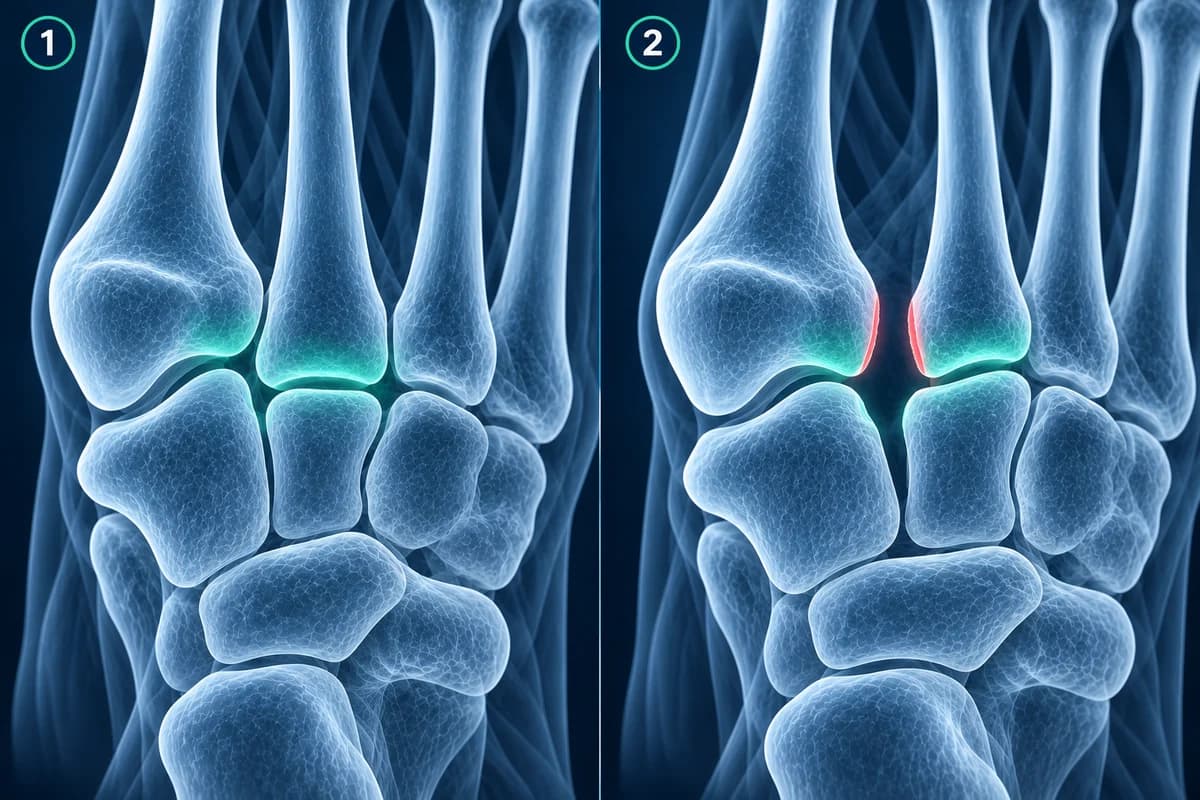
Start at the heel and work forward. On the side view the talus should sit squarely on the heel bone, and a line through the talus should continue straight down the first metatarsal; a sag in that line is a sign of flat foot. On the top-down view, the inner edge of the second metatarsal should line up with the inner edge of the middle cuneiform, and on the oblique view the inner edge of the fourth metatarsal should line up with the cuboid. A step at either spot, or a gap between the first and second metatarsal bases, raises concern for a Lisfranc injury.
In the forefoot, the big toe should point roughly along its metatarsal. When it drifts toward the second toe, the report measures the hallux valgus and intermetatarsal angles; our guide to foot X-ray angles explains each number and how a bunion is graded.
What does a foot fracture look like on X-ray?
A fresh fracture is a dark line or a step in the smooth white outer shell of a bone, called the cortex. In the foot the commonest places to look are the base of the fifth metatarsal, the shafts of the second and third metatarsals, the toes and the heel. Near the base of the fifth metatarsal, location decides treatment: a small piece pulled off the tip is usually an avulsion fracture, while a break slightly further along the bone is the slower-healing Jones fracture.
Overuse cracks behave differently. A metatarsal stress fracture is often invisible on the first film and only shows two to three weeks later as a faint line or a fluffy cuff of new bone around the shaft, which reports call “periosteal reaction” or “callus”. That is why a normal early X-ray does not end the story; read more in why a stress fracture may not show on X-ray. A heel bone crushed in a fall loses its normal upward angle on the side view, which is how a calcaneal fracture is often first recognised.
What do common foot X-ray report phrases mean?
- “Fleck sign”: a tiny bone chip between the first and second metatarsal bases, often the Lisfranc ligament pulling off a piece of bone. It is small but important.
- “Accessory ossicle” or “os naviculare / os peroneum”: an extra small bone many people are born with. Its edges are smooth and rounded, unlike the sharp edges of a fresh fracture.
- “Bipartite sesamoid”: one of the two pea-shaped bones under the big toe joint formed in two parts from birth, a frequent look-alike for a sesamoid fracture.
- “Joint space narrowing with dorsal osteophyte”: wear of the big toe joint with a bone spur on top, the classic picture of hallux rigidus.
- “Punched-out erosions with overhanging edges”: bitten-looking defects near a joint that suggest long-standing gout.
- “Soft tissue swelling, no acute osseous abnormality”: the bones look intact on this film. It does not rule out ligament, tendon or early stress injury.
What does arthritis or a deformity look like on a foot X-ray?
Wear-and-tear arthritis narrows the gap between bones and grows spurs at their edges; at the big toe this is hallux rigidus. Inflammatory arthritis tends to thin the bone around the joints and erode the metatarsal heads, while gout leaves sharper, overhanging bites, described in gout in the foot. A flattened, irregular second metatarsal head in a teenager or young adult points to Freiberg disease. In people with diabetes and reduced feeling, a midfoot that looks fragmented and collapsed raises the question of Charcot foot, which needs prompt specialist care.
What can a foot X-ray miss?
X-rays show bone well but soft tissue poorly. Plantar fascia, tendons, the plantar plate under the toe joints and nerves such as a Morton’s neuroma are essentially invisible. Early stress injuries, bone bruises and some subtle midfoot sprains can also hide on a normal-looking film. When pain continues, the next test is usually MRI, ultrasound or CT, chosen by the question being asked; our comparison of foot MRI, X-ray and ultrasound walks through which one fits which problem.
When is foot pain an emergency?
- You cannot put weight on the foot, or it looks bent or out of place
- A toe or the foot turns pale, blue or cold, or loses feeling
- Bone is visible, or there is an open wound over a possible fracture
- A hot, red, swollen foot with diabetes, especially with fever or a wound that will not heal
These need same-day urgent care whatever the X-ray report says. A report that mentions gas in the soft tissues of a diabetic foot is also an emergency.
Can I look at my own foot X-ray images?
Yes. Most imaging centres give you a disc or download link with DICOM files. Open them in our free X-ray viewer, which runs in your browser without uploading the original files, to zoom into the metatarsal bases or compare both feet side by side. Looking for yourself is a way to follow your report, not to replace the radiologist.
Frequently Asked Questions
Why was my foot X-ray normal if it still hurts?
X-rays show bone, not ligaments, tendons, nerves or the plantar fascia, and early stress fractures often do not appear for two to three weeks. If pain persists, a repeat X-ray, MRI or ultrasound may be considered by your clinician.
Why did they X-ray both of my feet?
Comparing the injured foot with the healthy one helps spot small differences, such as a slight widening between the first and second metatarsal bases or a normal growth plate in a child that could be mistaken for a fracture.
What is the difference between an accessory bone and a fracture fragment?
An accessory bone is usually rounded with a smooth white rim on every side and often appears in both feet. A fracture fragment tends to have a sharp, uneven edge and a matching defect on the parent bone. When it is unclear, the radiologist may suggest comparison views or MRI.
Does a weight-bearing foot X-ray matter?
Yes, for alignment questions. Bunion angles, arch height and subtle Lisfranc widening are judged on standing films, because the foot changes shape under load. For a fresh injury where standing hurts too much, the first films may be taken lying down.
Can AI read my foot X-ray?
AI can describe visible patterns and translate report wording into plain language, but it is not a radiologist and cannot examine your foot. Use it to prepare questions, then review the result with a clinician.
Sources
- Expert Panel on Musculoskeletal Imaging; Gorbachova T, Chang EY, Ha AS, et al. ACR Appropriateness Criteria Acute Trauma to the Foot. J Am Coll Radiol. 2020;17(5S):S2-S11.
- Expert Panel on Musculoskeletal Imaging; Said N, Fox MG, Nacey NC, et al. ACR Appropriateness Criteria Chronic Foot Pain: Update 2025. J Am Coll Radiol. 2026;23(4):681-699.
- Stiell IG, Greenberg GH, McKnight RD, et al. Decision rules for the use of radiography in acute ankle injuries. Refinement and prospective validation. JAMA. 1993;269(9):1127-1132.
- Bachmann LM, Kolb E, Koller MT, et al. Accuracy of Ottawa ankle rules to exclude fractures of the ankle and mid-foot: systematic review. BMJ. 2003;326(7386):417.
Related Articles
What the numbers on your foot X-ray mean: hallux valgus angle, intermetatarsal angle, Meary's angle, calcaneal pitch and Böhler's angle, with normal ranges and how each is measured.
X-ray, MRI, ultrasound or CT for foot pain? What each test shows and misses, which one usually comes first, and how the choice changes for stress fractures, neuromas, plantar fasciitis, Lisfranc injuries and the diabetic foot.
Why Lisfranc midfoot injuries are missed on first X-rays, the alignment lines and fleck sign to check, and when standing X-rays, CT or MRI are needed.
Related Conditions
Understand your imaging before your next appointment
MRI, X-ray or CT from a CD, folder or ZIP. A PDF report or photos of the films work too.
Free to open and preview, no account needed. An optional AI report from $10 explains your images in plain language, with questions for your doctor.
Your raw files stay on this device. Only rendered images and minimal context are sent when you request an AI report.
Medical Disclaimer: This page is for informational and educational purposes only. It does not constitute medical advice, diagnosis, or treatment. AI-generated analysis may contain errors. Always consult a qualified healthcare professional for medical decisions. Full Disclaimer