
Calcaneal (Heel Bone) Fracture: Böhler Angle, CT Findings and Recovery
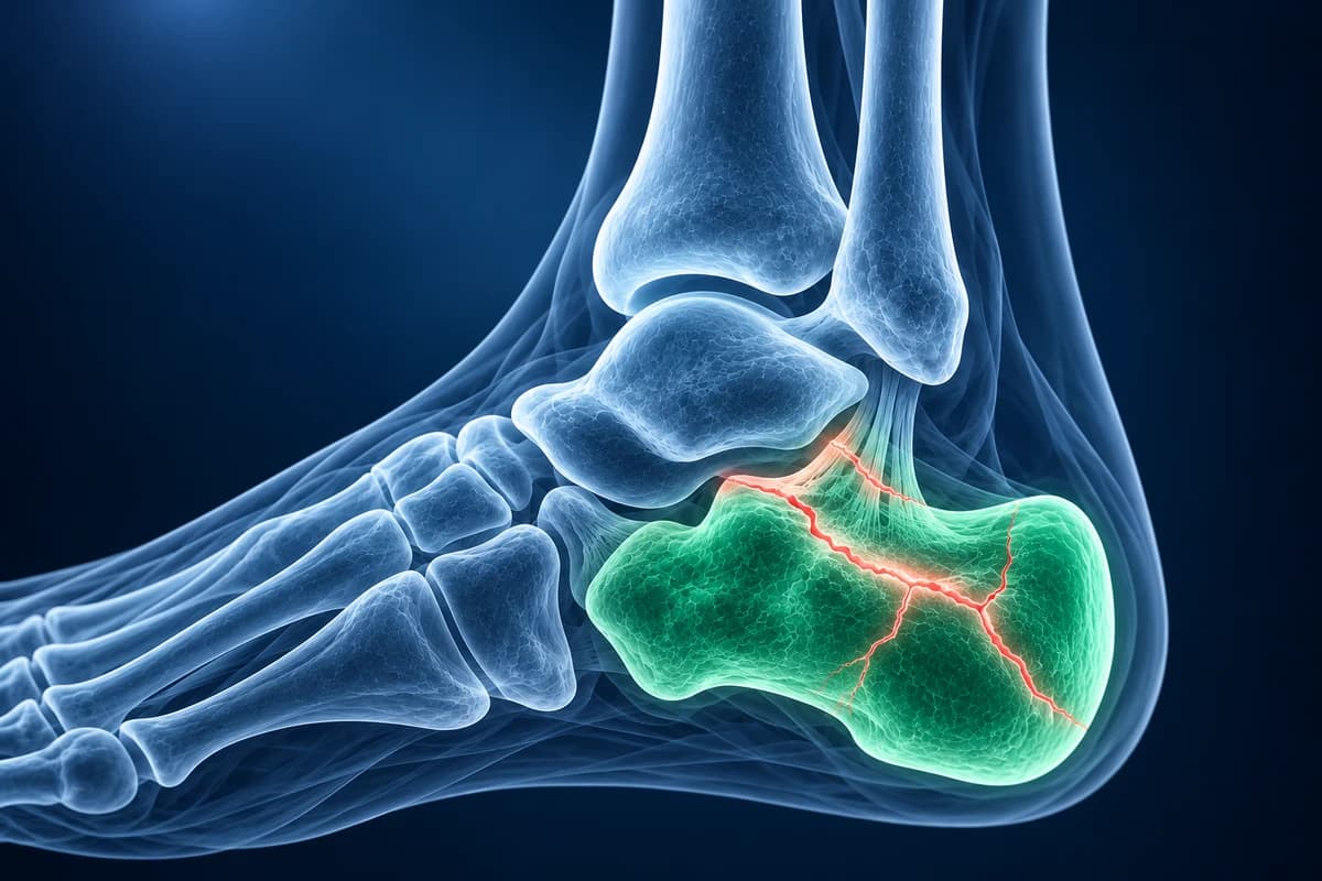
A calcaneal fracture is a break in the heel bone, usually from landing on the feet after a fall. On a side X-ray, doctors measure Böhler's angle, normally about 20° to 40°; a lower value means the top of the heel bone has been crushed down. CT then shows how many pieces the joint surface is broken into, which guides treatment.
Some signs described on this page need prompt medical care. If you have them, get care first. Reading about imaging can wait.
Common Symptoms
- Severe heel pain right after a fall from height, a jump or a car crash in which the foot hit the floor pedal
- Inability to put weight on the heel
- Rapid swelling that widens the heel, often with bruising that spreads to the sole and arch
- Fluid-filled blisters on the skin of the heel or ankle in the first days after injury
- Pain on squeezing the heel from both sides, a common finding in calcaneal stress fractures in runners
- Later stiffness and pain when walking on uneven ground, reflecting damage to the subtalar joint
Which heel fracture symptoms need emergency care?
A heel that cannot take weight after a fall needs same-day emergency assessment. Act immediately if pain is severe and rising with a tense, swollen foot, if stretching the toes hurts sharply, or if the foot goes numb, pale or cold: these are warning signs of compartment syndrome. Skin that is broken, or skin over the back of the heel that is pale, tight and pushed outward by a bone fragment, is a surgical emergency because the skin can die. Because the same fall often injures the spine, back pain after a heel fracture also needs to be mentioned to the emergency team.
How does the heel bone break?
The calcaneus is the largest bone in the foot. Its upper surface carries the talus at the subtalar joint, the joint that lets the heel tilt in and out on uneven ground. In a fall onto the heel, the talus is driven down like a wedge into the calcaneus, splitting it and pushing the joint surface into the soft bone beneath. Most fractures from this mechanism involve the subtalar joint (intra-articular) and are the ones that most affect long-term function.
Smaller extra-articular fractures also happen: a chip off the front of the bone (anterior process) during a twist, which is often first labelled an ankle sprain; an avulsion where the Achilles tendon pulls off part of the back of the heel, which is managed alongside the tendon; and stress fractures in runners and military recruits, which cause gradual heel pain that can be confused with plantar fasciitis. Compare symptoms resembling a calcaneal stress fracture in our heel-pain guide.
What is Böhler's angle and why is mine reduced?
Böhler's angle is drawn on the side X-ray using two lines: one from the front top of the calcaneus to the highest point of the posterior joint surface, and one from that point to the top of the heel tuberosity at the back. In a normal heel, the angle between them is roughly 20° to 40°. When the joint surface is driven down, the middle point drops and the angle flattens or even becomes negative. A reduced angle points to a depressed joint fragment, but a normal angle does not exclude a fracture, so the full images and examination still count. Our guide to foot X-ray angles shows how this and related measurements are drawn.
Reports may also mention the critical angle of Gissane, formed by the edges of the bone just beneath the talus. It tends to widen when the joint surface collapses. Both angles are helpful for comparing before and after treatment. Review the surrounding terminology in the foot X-ray report guide when reading heel measurements.
Why is CT needed for a calcaneal fracture?
Plain films usually include a side view, an axial (Harris) view looking up at the heel from behind, and views of the rest of the foot. They confirm the fracture but cannot show the joint surface well because the bone is irregular and overlaps itself. The ACR Appropriateness Criteria for acute foot trauma support CT when radiographs show a complex hindfoot fracture or when a fracture is suspected despite normal films.
Surgeons widely use the Sanders classification, based on coronal CT through the widest part of the posterior facet. Type I is undisplaced (less than 2 mm). Types II, III and IV describe the joint surface broken into two, three, or four or more displaced pieces. Sanders and colleagues reported that outcomes worsen as the type rises. CT also shows widening of the heel, fragments pressing on the peroneal tendons and involvement of the joint with the cuboid. MRI is reserved for suspected stress fractures or bone bruising when X-rays are normal. You can open a heel CT in the free CT viewer.
What do calcaneal fracture report phrases mean?
- "Intra-articular fracture with posterior facet depression"
- The break reaches the main joint under the talus and part of that joint surface has been pushed down.
- "Joint depression type" vs "tongue-type"
- Two common patterns. In a tongue-type fracture, the joint fragment stays attached to a long piece of the back of the heel, which can tilt up and press on the skin.
- "Decreased Böhler angle"
- The top of the heel bone has collapsed compared with a normal heel profile.
- "Lateral wall blowout" or "heel widening"
- The outer wall of the heel has bulged out, which can crowd the peroneal tendons and make shoes hard to wear.
- "Extension to the calcaneocuboid joint"
- The fracture also reaches the joint at the front of the heel bone, adding a second joint injury.
For other X-ray wording, see how to read a foot X-ray.
What we look for on heel imaging
- Böhler's angle and Gissane's angle on the lateral view, compared with normal ranges
- Fracture lines entering the posterior facet of the subtalar joint
- Number and displacement of joint fragments on coronal CT
- Heel widening, shortening, varus tilt and lateral wall bulging on the axial view
- Extension into the calcaneocuboid joint or the anterior process
- Fine sclerotic bands or marrow edema of a calcaneal stress fracture
Does a broken heel need surgery, and how long is recovery?
Undisplaced and extra-articular fractures are usually treated without surgery: rest, elevation, a boot or cast and no weight on the heel for several weeks, with early ankle and toe movement. Displaced intra-articular fractures may be fixed with plates and screws, sometimes through small incisions, to restore the joint and heel shape. Surgery is timed around the swelling, because operating through swollen, blistered skin raises the risk of wound problems. Smoking and diabetes increase that risk, and in some people non-operative care is the safer choice even when the joint is displaced.
Recovery is long either way. Many people are off the foot for six to eight weeks or more, and it can take a year or longer to reach the final result. Stiffness of the subtalar joint is common, and some develop arthritis that may later need a fusion. Your surgeon will explain how your CT type affects the options.
Can I review my own heel X-ray or CT?
Open your images privately in the free foot X-ray viewer to look at the heel profile, or upload them for a plain-language AI explanation of the angles and report terms. The explanation is educational and not a diagnosis; your orthopaedic team makes the treatment decisions. If a stiff, painful hindfoot has troubled you since childhood, a tarsal coalition or flat foot may also be worth discussing.
Frequently Asked Questions
What is a normal Böhler angle?
Böhler's angle is usually between about 20° and 40° on a lateral X-ray. Values below that range suggest the joint surface has collapsed. Because normal values vary between people, an X-ray of the uninjured heel is sometimes used for comparison.
Can you walk on a fractured calcaneus?
Most people cannot walk on an acute calcaneal fracture, and treatment usually involves weeks without weight on the heel. Small stress fractures may allow walking with pain, but they still need assessment. Do not test the heel by forcing weight onto it.
Why did the hospital also X-ray my back?
A fall hard enough to break the heel can also compress a vertebra, most often in the lower back, and can break the other heel. Teams therefore examine the spine and both feet, and image them if there is tenderness or the mechanism suggests it.
What does Sanders type III mean?
Sanders type III means CT shows the main joint surface under the talus broken into three displaced pieces. It is more complex than type II and is often considered for surgical repair, but the final decision depends on your health, skin condition and activity goals.
Will my heel ever feel normal again?
Many people return to walking and work, but some stiffness or ache on uneven ground is common after intra-articular fractures. Simpler fracture patterns tend to do better. Physiotherapy, supportive footwear and, where needed, later surgery for arthritis can help.
Sources
- Sanders R, Fortin P, DiPasquale T, Walling A. Operative treatment in 120 displaced intraarticular calcaneal fractures. Results using a prognostic computed tomography scan classification. Clin Orthop Relat Res. 1993;(290):87-95.
- Seaman TJ, Bergman R. Calcaneus Fractures. StatPearls. Treasure Island (FL): StatPearls Publishing.
- Expert Panel on Musculoskeletal Imaging; Gorbachova T, Chang EY, Ha AS, et al. ACR Appropriateness Criteria Acute Trauma to the Foot. J Am Coll Radiol. 2020;17(5S):S2-S11.
- American Academy of Orthopaedic Surgeons. Calcaneus (Heel Bone) Fractures. OrthoInfo.
Related Articles
How long plantar fasciitis lasts, what changes month by month, why morning pain is the last to go, and when imaging or a different diagnosis should be considered.
Heel pain mapped to its causes: plantar fasciitis, heel stress fracture, Baxter's nerve, fat pad atrophy and Sever's disease, and which test shows each one.
What the numbers on your foot X-ray mean: hallux valgus angle, intermetatarsal angle, Meary's angle, calcaneal pitch and Böhler's angle, with normal ranges and how each is measured.
Do not delay urgent medical care to use this tool.
After you get care, understand your imaging
MRI, X-ray or CT from a CD, folder or ZIP. A PDF report or photos of the films work too.
Free to open and preview, no account needed. An optional AI report from $10 explains your images in plain language, with questions for your doctor.
Your raw files stay on this device. Only rendered images and minimal context are sent when you request an AI report.
Medical Disclaimer: This page is for informational and educational purposes only. It does not constitute medical advice, diagnosis, or treatment. AI-generated analysis may contain errors. Always consult a qualified healthcare professional for medical decisions. Full Disclaimer