
Heel Pain Causes and Imaging: Which Test Shows What
Heel pain mapped to its causes: plantar fasciitis, heel stress fracture, Baxter's nerve, fat pad atrophy and Sever's disease, and which test shows each one.
Key Takeaways
- Most pain under the heel is plantar fasciitis and is diagnosed without a scan
- Pain when both sides of the heel are squeezed suggests a stress fracture, which MRI shows earlier than X-ray
- Baxter’s nerve entrapment shows on MRI as fat replacing a small muscle on the outer sole
- A heel spur is common in people without pain and does not prove the cause
- Back-of-heel pain belongs with the Achilles tendon; in children it is often Sever’s disease
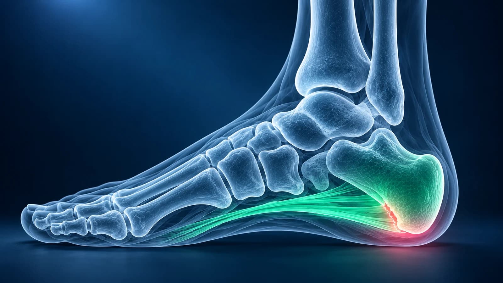
Pain under the heel is usually plantar fasciitis, and it is normally diagnosed from your history and an examination without any scan. Imaging is added when the pain lasts for months, the pattern does not fit, or a child is affected: an X-ray first, then ultrasound or MRI. On ultrasound, a plantar fascia thicker than 4 mm supports plantar fasciitis.
This hub lists the common causes of pain under and around the heel, shows which test answers which question, and sends you to the detailed page for each cause. It is educational and does not replace an examination by a clinician who can press on the right spots.
When does heel pain need to be seen quickly?
- You cannot put weight on the heel after a fall from a height or a car accident, which can break the heel bone.
- The heel is hot, red and swollen, or you have a fever, especially with diabetes or a wound on the foot.
- You felt a pop at the back of the heel and now struggle to push off, which can mean an Achilles tendon rupture.
- Numbness spreading across the sole, or pain that wakes you at night and does not ease with rest.
Which test shows which cause of heel pain?
The ACR Appropriateness Criteria for chronic foot pain start almost every heel problem with plain X-rays and reserve ultrasound or MRI for the questions X-ray cannot answer. The table pairs each cause with the test that usually settles it.
| Cause | Where it hurts | Test that shows it best | Typical report wording |
|---|---|---|---|
| Plantar fasciitis | Inner front edge of the heel, worst on the first steps | Examination; ultrasound or MRI if it persists | Thickened proximal plantar fascia, perifascial edema |
| Heel bone stress fracture | Both sides of the heel when squeezed, after more running or walking | MRI; X-ray is often normal early on | Calcaneal marrow edema with a low-signal fracture line |
| Baxter’s nerve entrapment | Inner heel, sometimes burning, may not ease after the first steps | MRI | Fatty atrophy of the abductor digiti minimi muscle |
| Heel fat pad atrophy | Centre of the heel, like walking on bone, worse on hard floors | Examination; ultrasound measures the pad | Thinned heel pad, reduced heel pad thickness |
| Sever’s disease (children) | Back and sides of the heel in an active child | Examination; X-ray only if the story is unusual | Sclerotic or fragmented calcaneal apophysis, which does not prove the diagnosis |
Where exactly is your heel pain?
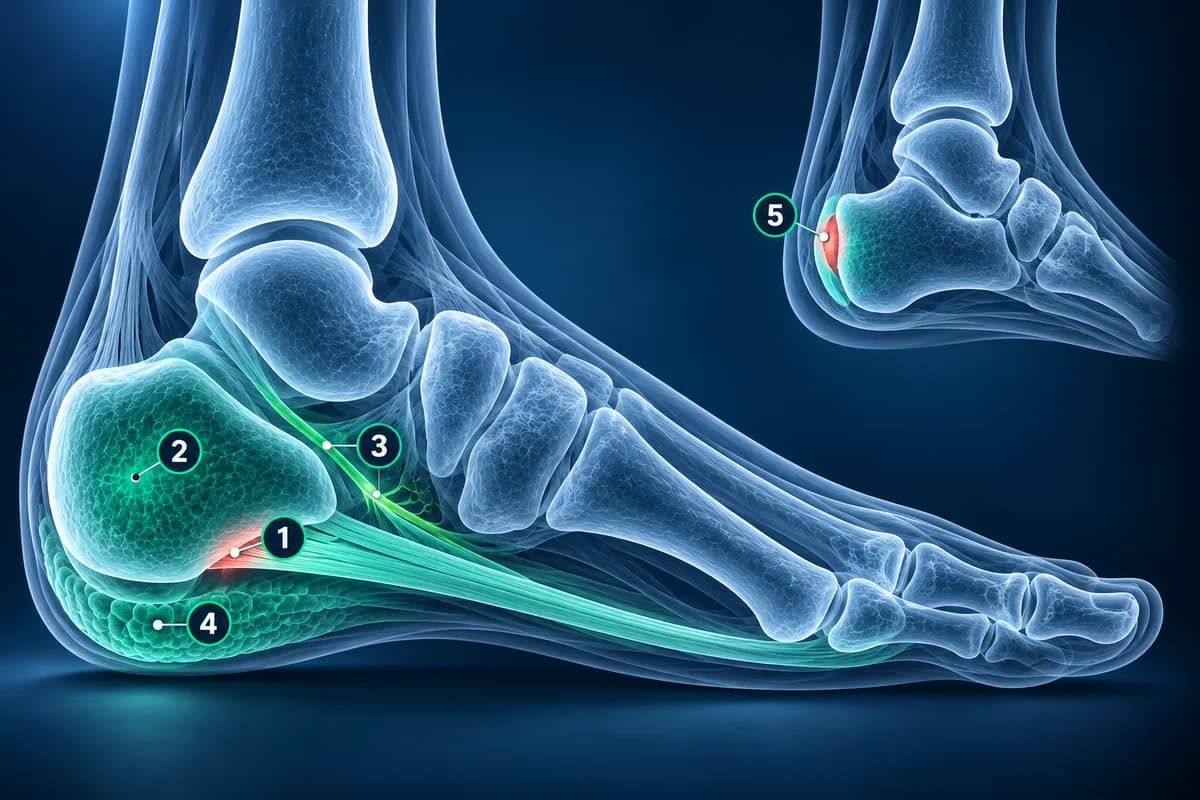
Under the front of the heel and slightly to the inner side points to the fascia. Pain when both sides of the heel are squeezed points to the bone. Pain over the middle of the pad, which feels like a bruise, points to the fat pad. Pain at the very back of the heel, above the sole, is a different family of problems that involves the Achilles tendon.
Is it plantar fasciitis?
Sharp pain with the first steps out of bed or after sitting, easing as you warm up and returning after long standing, is the classic pattern of plantar fasciitis. The 2023 physical therapy clinical practice guideline treats it as a clinical diagnosis. Ultrasound is useful when the diagnosis is uncertain: a fascia thicker than 4 mm at the heel, darker than normal and sometimes with fluid around it supports the diagnosis. Whether that justifies an MRI is covered in do you need an MRI for plantar fasciitis.
A heel spur on the X-ray is a frequent source of worry. Spurs are common in people with heel pain but are also seen in people without any pain, so a spur on its own does not explain the symptoms and does not need to be removed.
Could it be a stress fracture of the heel bone?
A calcaneal stress fracture follows a jump in training, a new walking routine or, in older adults, thinning bone. The tell-tale sign is pain when the clinician squeezes both sides of the heel together. X-rays taken in the first weeks are often normal; a faint white band of healing bone may appear only once healing starts, several weeks later. MRI shows the bright band of marrow edema with a dark line through it much earlier. Read why in stress fracture not on X-ray. A heel broken by a fall from a height is a different injury, covered on the calcaneal fracture page.
What is Baxter’s nerve entrapment?
Baxter’s nerve is a small branch of the lateral plantar nerve that runs under the inner heel to the muscle that moves the little toe. When it is squeezed, the pain sits slightly further back and more to the inner side than plantar fasciitis and may burn. It is easy to miss because it often coexists with a thick fascia. On MRI the clue is indirect: the abductor digiti minimi muscle on the outer sole has been replaced by fat. Fluid-sensitive images open in our free MRI viewer if you want to see the slices the report describes.
What does “heel fat pad atrophy” mean?
The heel pad is a honeycomb of fat chambers that absorbs each step. With age, steroid injections or years of hard impact it can thin and lose its spring, and the pain feels like a deep bruise in the centre of the heel rather than at the front edge. Ultrasound can measure the thickness of the pad and compare it with the other foot. The report may call it “fat pad atrophy” or “heel pad contusion” when the pad is bruised after a landing.
Why does my child’s heel hurt?
In children who are still growing, the heel bone has a growth plate at the back where the Achilles tendon pulls. Running and jumping sports can irritate it, which is Sever’s disease. The diagnosis is clinical. On X-ray a dense heel growth centre is normal in children without pain, and neither a dense nor a fragmented growth centre proves the diagnosis, so when an X-ray is taken it is to look for another cause such as a bone cyst or stress fracture. More childhood foot causes are gathered in children’s foot pain and imaging.
What if the pain is at the back of the heel or burns into the sole?
Pain where the Achilles tendon attaches to the back of the heel is assessed as a tendon problem; our ankle site explains Achilles tendon tears and insertional tendinopathy.
A bony bump at the back of the heel that rubs on shoes, with a swollen bursa in front of the tendon, is a Haglund deformity, which the same Achilles page on the ankle site covers alongside the tendon.
Burning, tingling or numbness spreading across the sole from behind the inner ankle suggests tarsal tunnel syndrome, a nerve problem assessed at the ankle.
What can a heel X-ray show, and what can it not?
A heel X-ray, usually a side view plus an angled view from below, shows spurs, fractures, the shape of the heel bone, arthritis of the joint below the ankle and, in children, the growth plate. It cannot show the fascia, nerves or fat pad directly. You can open your own study in our free X-ray viewer and follow the steps in how to read a foot X-ray.
Frequently Asked Questions
What is the most common cause of heel pain?
In adults it is plantar fasciitis, an overload of the fascia where it attaches to the heel bone. In active children who are still growing it is Sever’s disease, an irritation of the heel growth plate.
Should I get an X-ray or an MRI for heel pain?
Most heel pain needs neither at first. If imaging is needed, an X-ray comes first to look at the bone. Ultrasound or MRI follow when the pain persists despite treatment or a stress fracture or nerve problem is suspected.
Does a heel spur cause heel pain?
Not usually on its own. Most people with a heel spur have no heel pain at all. When there is pain, it usually comes from the plantar fascia, not from the spur itself.
Can an ultrasound diagnose plantar fasciitis?
It can support the diagnosis. A fascia thicker than 4 mm near the heel, with darker, swollen fibres, fits plantar fasciitis, and ultrasound also guides injections. A normal ultrasound with typical symptoms does not rule it out completely.
Can AI tell me what is causing my heel pain?
AI can explain what an X-ray, ultrasound or MRI report says about each heel structure and suggest questions for your appointment. The cause of heel pain is decided by a clinician who has examined your foot.
Sources
- Expert Panel on Musculoskeletal Imaging; Said N, Fox MG, Nacey NC, et al. ACR Appropriateness Criteria Chronic Foot Pain: Update 2025. J Am Coll Radiol. 2026;23(4):681-699.
- Koc TA Jr, Bise CG, Neville C, et al. Heel Pain - Plantar Fasciitis: Revision 2023. J Orthop Sports Phys Ther. 2023;53(12):CPG1-CPG39.
- McMillan AM, Landorf KB, Barrett JT, Menz HB, Bird AR. Diagnostic imaging for chronic plantar heel pain: a systematic review and meta-analysis. J Foot Ankle Res. 2009;2:32.
- Bergman R, Kaiser K. Stress Reaction and Fractures. StatPearls. Treasure Island (FL): StatPearls Publishing.
- Chundru U, Liebeskind A, Seidelmann F, et al. Plantar fasciitis and calcaneal spur formation are associated with abductor digiti minimi atrophy on MRI of the foot. Skeletal Radiol. 2008;37(6):505-510.
- American Academy of Orthopaedic Surgeons. Plantar Fasciitis and Bone Spurs. OrthoInfo.
- Volpon JB, de Carvalho Filho G. Calcaneal apophysitis: a quantitative radiographic evaluation of the secondary ossification center. Arch Orthop Trauma Surg. 2002;122(6):338-341.
- Kose O. Do we really need radiographic assessment for the diagnosis of non-specific heel pain (calcaneal apophysitis) in children? Skeletal Radiol. 2010;39(4):359-361.
Related Articles
Most plantar fasciitis needs no MRI. Learn when an X-ray, ultrasound or MRI is worth it, what each shows, and what heel pain report phrases mean.
Why a foot stress fracture can hide on a normal X-ray, what MRI, CT and repeat X-rays show instead, and which foot stress fractures are high risk.
Why a child's foot hurts: Sever's, Köhler, tarsal coalition, accessory navicular and toddler fractures, what X-rays show and the red flags to act on.
Related Conditions
Understand your imaging before your next appointment
MRI, X-ray or CT from a CD, folder or ZIP. A PDF report or photos of the films work too.
Free to open and preview, no account needed. An optional AI report from $10 explains your images in plain language, with questions for your doctor.
Your raw files stay on this device. Only rendered images and minimal context are sent when you request an AI report.
Medical Disclaimer: This page is for informational and educational purposes only. It does not constitute medical advice, diagnosis, or treatment. AI-generated analysis may contain errors. Always consult a qualified healthcare professional for medical decisions. Full Disclaimer