
Children's Foot Pain: Causes, X-rays and When to Worry
Why a child's foot hurts: Sever's, Köhler, tarsal coalition, accessory navicular and toddler fractures, what X-rays show and the red flags to act on.
Key Takeaways
- Fever, refusing to walk and night pain in a child need same-day medical review
- Sever’s disease is the usual cause of heel pain in active children and is diagnosed by examination
- Köhler disease makes the navicular look flat and dense, but it usually heals on its own
- A stiff flat foot in a teen suggests tarsal coalition, confirmed on oblique X-ray, CT or MRI
- Growth centres and extra bones are smooth and rounded; fractures are sharp-edged and tender
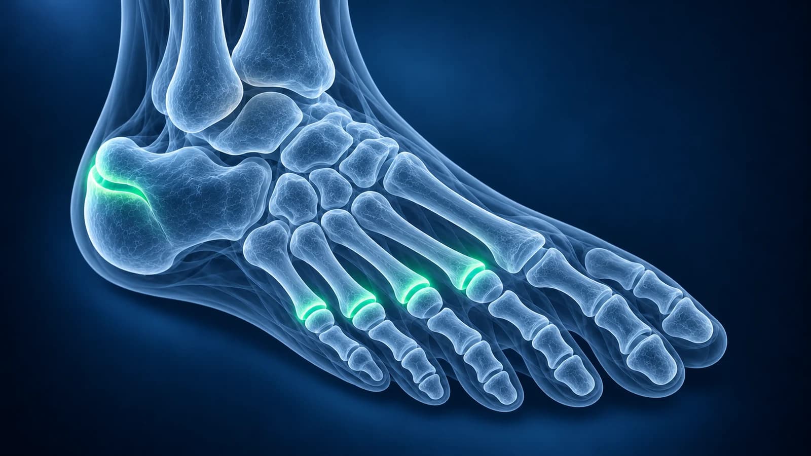
When does a child with foot pain need to be seen the same day?
- Fever, or a foot or ankle that is hot, red and swollen, which can mean a bone or joint infection
- Refusing to stand or walk, or a new limp in a young child, even without a known fall
- Pain that wakes the child at night, gets steadily worse or comes with weight loss or tiredness
- A visibly bent foot or toe after an injury, or a child who seems unwell
Most foot pain in active children comes from overloaded growth areas: the heel in Sever’s disease, the navicular in Köhler disease or an extra bone on the inner arch. These are usually diagnosed by examination and often need no X-ray. Fever, refusing to walk or night pain change that and need prompt medical review.
Children’s X-rays are harder to read than adults’ because growth plates, separate growth centres and extra bones are everywhere. This hub explains the common causes, which test answers which question and which normal shapes are often mistaken for fractures. It is educational and does not replace an examination.
Which test shows which cause of foot pain in a child?
For a child up to age 5 with a new limp, the ACR Appropriateness Criteria start with radiographs of the area that hurts when infection is not suspected, and move to MRI, or to ultrasound when the hip is the source, when infection is a concern. Children should receive as little radiation as possible, so the examination decides whether an X-ray is needed at all.
| Cause | Typical child | Test that shows it best | Typical report wording |
|---|---|---|---|
| Sever’s disease | Sporty child aged about 8 to 15, heel pain after running | Examination; X-ray use varies | Sclerotic, fragmented calcaneal apophysis |
| Köhler disease | Young child limping, pain in the middle of the inner arch | X-ray | Flattened, dense or fragmented navicular |
| Accessory navicular | Older child or teen, tender bump on the inner arch | X-ray; MRI if painful and unclear | Accessory navicular, edema across the synchondrosis |
| Tarsal coalition | Teen with a stiff flat foot or repeated ankle sprains | X-ray with oblique view; CT or MRI to confirm | Calcaneonavicular or talocalcaneal coalition, anteater nose sign, C-sign |
| Toddler’s foot fracture | Toddler refusing to walk after a minor fall or none remembered | X-ray, repeated later if normal | Subtle sclerosis or periosteal reaction in the cuboid, calcaneus or a metatarsal |
Which growth areas in a child’s foot cause pain?
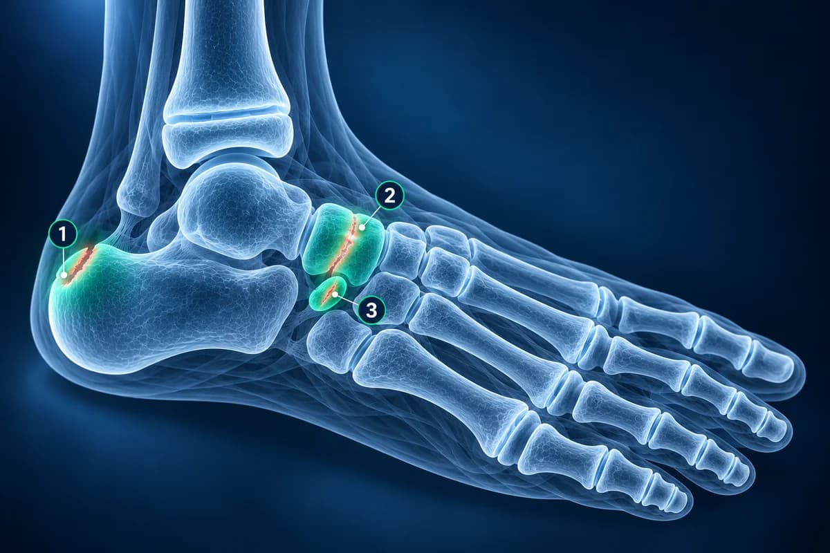
Why does my child’s heel hurt after sport?
The back of the heel bone grows from a separate cap of bone called the apophysis, where the Achilles tendon pulls. During growth spurts, running and jumping irritate it, which is Sever’s disease. Squeezing the back of the heel from both sides hurts, and both heels can be affected. A dense apophysis can be normal in children without pain, so it does not prove the diagnosis, and many clinicians skip the X-ray in typical cases. Others take one routinely, because in one series about 1 in 20 children diagnosed with Sever’s disease turned out to have another cause, such as a bone cyst or a stress fracture. Adult heel causes are compared in heel pain causes and imaging.
What does a flattened navicular on a child’s X-ray mean?
In Köhler disease the navicular bone in the middle of the arch temporarily loses part of its blood supply. On X-ray it looks thin, white and sometimes broken into pieces. The appearance is alarming, but the bone usually rebuilds on its own, and treatment aims at comfort, sometimes with a short period in a walking cast. An irregular navicular can also be found in a child without pain, so the X-ray is always read together with the symptoms.
What is the bump on the inner side of my child’s foot?
An accessory navicular is an extra bone next to the navicular, where the tendon that supports the arch attaches. It is common and usually painless, but in older children and teens the junction between the two bones can become irritated by sport or tight shoes. The X-ray shows the extra bone; MRI shows whether the junction is inflamed. It is sometimes mistaken for a chip fracture.
Could a stiff flat foot in a teenager be a tarsal coalition?
A tarsal coalition is a bar of bone, cartilage or fibrous tissue joining two bones of the back of the foot. It is present from birth but tends to cause pain in early adolescence, when the bar stiffens. The arch stays flat even on tiptoe and ankle sprains keep recurring. An oblique X-ray shows a bar between the heel bone and the navicular, while a coalition under the ankle may need CT or MRI. A flexible flat foot that forms an arch on tiptoe is different and usually harmless; see flat foot.
Can a toddler break a foot bone without a clear fall?
Yes. Toddlers can crack the cuboid, the heel bone or a metatarsal from a jump off a sofa or a twist on a step, and then limp or refuse to walk. The first X-ray is often normal; a repeat X-ray later can show a faint white line of healing bone. Because a limping toddler can also have an infection, fever or a child who seems unwell always needs prompt review. Our article on fractures that do not show on X-ray explains why early films can miss these.
Which normal growth plates and extra bones look like fractures?
Several normal structures in a child’s foot are regularly mistaken for breaks. The growth centre at the base of the fifth metatarsal runs parallel to the bone, which helps tell it from a fracture. Extra bones such as the accessory navicular and the os peroneum have smooth, rounded edges, while a fresh fracture fragment has sharp edges and matching swelling. The heel apophysis looks dense in pain-free children too. When in doubt, radiologists compare with the other foot or with the expected appearance for the child’s age. You can look at the images yourself in our free X-ray viewer alongside how to read a foot X-ray.
Frequently Asked Questions
Is foot pain in children just growing pains?
Classic growing pains are aching in both legs in the evening or night that is gone by morning and does not cause a limp. Pain in one specific spot of the foot, a limp or swelling points to a specific cause and should be examined.
Does my child need an X-ray for heel pain?
Not always. Sever’s disease is diagnosed by examination, and an X-ray cannot prove it. Some clinicians still take one routinely because about 1 in 20 children with typical symptoms turned out to have another cause in one series. Pain at rest or at night, swelling, or pain that does not settle with rest from sport should always be examined.
Can a growth plate be mistaken for a fracture on X-ray?
Yes, and the reverse also happens. Growth plates and extra bones have smooth edges and typical locations, so radiologists compare their shape and direction with known normal patterns and sometimes with the other foot.
Why would a child need an MRI of the foot?
MRI is used when infection is suspected, when a fracture is likely but the X-ray is normal, to confirm a tarsal coalition or to see whether an accessory bone is inflamed. It uses no radiation but young children may need sedation to keep still.
Can AI explain my child’s foot X-ray report?
AI can explain terms such as apophysis, ossicle or sclerosis in plain language before your appointment. It does not examine your child, and a limping child with fever needs a clinician, not an app.
Sources
- Expert Panel on Pediatric Imaging; Safdar NM, Rigsby CK, Iyer RS, et al. ACR Appropriateness Criteria Acutely Limping Child Up To Age 5. J Am Coll Radiol. 2018;15(11S):S252-S262.
- Lawrence DA, Rolen MF, Haims AH, Zayour Z, Moukaddam HA. Tarsal Coalitions: Radiographic, CT, and MR Imaging Findings. HSS J. 2014;10(2):153-166.
- Vora BMK, Wong BSS. Common accessory ossicles of the foot: imaging features, pitfalls and associated pathology. Singapore Med J. 2018;59(4):183-189.
- American Academy of Orthopaedic Surgeons. Tarsal Coalition. OrthoInfo.
- Rachel JN, Williams JB, Sawyer JR, Warner WC, Kelly DM. Is radiographic evaluation necessary in children with a clinical diagnosis of calcaneal apophysitis (Sever disease)? J Pediatr Orthop. 2011;31(5):548-550.
- Smith JM, Varacallo MA. Sever Disease (Calcaneal Apophysitis). StatPearls. Treasure Island (FL): StatPearls Publishing.
- Williams GA, Cowell HR. Köhler’s disease of the tarsal navicular. Clin Orthop Relat Res. 1981;(158):53-58.
- John SD, Moorthy CS, Swischuk LE. Expanding the concept of the toddler’s fracture. Radiographics. 1997;17(2):367-376.
Related Articles
Heel pain mapped to its causes: plantar fasciitis, heel stress fracture, Baxter's nerve, fat pad atrophy and Sever's disease, and which test shows each one.
A plain-language guide to your foot X-ray: the three standard views, how to trace alignment from heel to toes, and what fractures, fleck signs, accessory bones and arthritis look like.
Why a foot stress fracture can hide on a normal X-ray, what MRI, CT and repeat X-rays show instead, and which foot stress fractures are high risk.
Related Conditions
Understand your imaging before your next appointment
MRI, X-ray or CT from a CD, folder or ZIP. A PDF report or photos of the films work too.
Free to open and preview, no account needed. An optional AI report from $10 explains your images in plain language, with questions for your doctor.
Your raw files stay on this device. Only rendered images and minimal context are sent when you request an AI report.
Medical Disclaimer: This page is for informational and educational purposes only. It does not constitute medical advice, diagnosis, or treatment. AI-generated analysis may contain errors. Always consult a qualified healthcare professional for medical decisions. Full Disclaimer