
Accessory Navicular: Normal Variant or Source of Pain?
An accessory navicular is an extra piece of bone on the inner side of the navicular, in the midfoot, where the arch-supporting posterior tibial tendon attaches. It is a common normal variant: a 2024 meta-analysis of 39 studies found it in about 12.6% of feet, and in half of affected people both feet have one. Most never hurt; pain usually comes from strain at the joint linking the extra bone to the navicular.
Common Symptoms
- A bony bump on the inner side of the midfoot, just in front of and below the ankle bone
- Redness, rubbing or a callus where shoes, ski boots or skates press on the bump
- Aching along the inner arch during or after running, jumping or long walks
- Tenderness directly on the bump, often starting in late childhood or the teenage years
- Pain that began after a twisting injury or a blow to the inner foot
- A flexible flat foot on the same side, with tiredness of the arch toward the end of the day
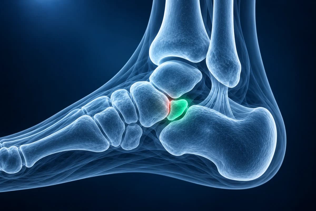
Radiology reports may also call it an os naviculare, os tibiale externum or a prehallux. Finding one on an X-ray taken for another reason is not a problem in itself. This page explains the three types, the MRI signs that suggest a symptomatic one, and how doctors tell it apart from a fracture. It is educational guidance, not a diagnosis.
What do type I, type II and type III accessory navicular mean?
- Type I: a small, round ossicle sitting inside the posterior tibial tendon, a few millimeters from the navicular and not connected to it. It behaves like a sesamoid bone and rarely causes symptoms.
- Type II: a larger, triangular or heart-shaped fragment joined to the navicular by a thin layer of cartilage or fibrous tissue, called a synchondrosis. Part of the tendon pulls on it, and repeated tugging or a twist can strain that link. This is the type most often painful.
- Type III: the extra bone has fused with the navicular, leaving a large, horn-like tuberosity called a cornuate navicular. It can hurt where shoes press on the prominence.
As the American College of Radiology criteria for chronic foot pain recommend, X-rays come first. The type is usually visible on a standing X-ray, best on a top-down view or an oblique view angled to show the inner side of the midfoot. You can look at your own views in the free X-ray viewer, and the foot X-ray reading guide shows where the navicular sits among the other bones.
What does “edema at the synchondrosis” mean on an MRI report?
On an X-ray, a painful and a painless accessory navicular can look the same. MRI shows whether the bone and its link are under stress. These are the phrases a report is likely to use. Use the foot X-ray report guide for accessory-navicular wording.
- “Bone marrow edema in the accessory navicular and adjacent navicular”: the bone on both sides of the link is swollen with fluid, a sign of ongoing stress. In a surgical study of painful type II ossicles by Choi and colleagues, every patient showed edema in the ossicle and the synchondrosis on fat-suppressed images.
- “Fluid signal at the synchondrosis” or “widening of the synchondrosis”: the cartilage or fibrous bridge looks bright or gapped, suggesting it has been strained or partly pulled apart.
- “Surrounding soft tissue edema” or “adventitial bursitis”: fluid in the tissue over the bump, often from shoe pressure.
- “Posterior tibial tendinosis at its navicular insertion”: the tendon is thickened or shows abnormal signal where it attaches. Tendon changes were common in the same study and matter for treatment planning.
- “No marrow edema, incidental accessory navicular”: the extra bone is present but quiet, so another cause of pain should be looked for.
A full walk-through of foot MRI sequences is in how to read a foot MRI, and MRI files can be opened in the free MRI viewer. If you are weighing whether the scan is worth it, see what a foot MRI costs.
Is it an accessory navicular or a fracture?
After an injury, a type II accessory navicular can be mistaken for a chip fracture of the navicular, and the reverse. An accessory bone has smooth, rounded, well-corticated edges and often a matching one in the other foot. A fresh fracture has sharp, irregular edges and fits back into the bone it came from. When the X-ray is unclear, MRI or CT can show whether the line is an old synchondrosis or a new break, and an X-ray of the other foot is sometimes taken for comparison.
Pain across the top of the midfoot after a twist is a different pattern and can mean a Lisfranc injury; it needs prompt assessment.
What We Look For on Imaging
- Size, shape and type of the ossicle beside the navicular tuberosity
- Width and regularity of the synchondrosis in a type II accessory navicular
- Bone marrow edema in the ossicle and the neighboring navicular on fluid-sensitive MRI
- Posterior tibial tendon thickening, signal change or tearing near its insertion
- Soft-tissue swelling or an inflamed bursa over the medial prominence
- Overall arch alignment and signs that separate an ossicle from an acute fracture
Does an accessory navicular cause flat feet?
The two often appear together, and older teaching held that the extra bone weakens the tendon's pull on the arch. The link is debated: many people with an accessory navicular have normal arches, and many flat feet have no extra bone. If your report also describes a low arch, the flat foot page explains the angles. In a teenager with a stiff rather than flexible flat foot, doctors also look for a tarsal coalition. Explore childhood foot-pain patterns alongside accessory-navicular symptoms.
A painful, swollen inner ankle with a sinking arch in an adult is often assessed as a posterior tibial tendon problem.
How is a painful accessory navicular treated, and when is it urgent?
Most painful accessory naviculars settle without surgery: softer or wider shoes, padding around the bump, an arch support, ice and a break from the sport that triggers it. A walking boot or cast for a few weeks may be used when pain is marked. If pain keeps returning over months, surgeons may remove the extra bone and reattach or tighten the posterior tibial tendon on the navicular, often called a Kidner-type procedure, or in some cases fuse the fragment to the navicular.
- Seek prompt care if you cannot bear weight after an injury or the midfoot is badly swollen and bruised.
- Seek same-day care for a hot, red, swollen foot with fever, an open sore over the bump, or any new red, warm swelling of the foot if you have diabetes.
Frequently Asked Questions
Is an accessory navicular a normal finding?
Yes. It is one of the most common extra bones in the foot, present in roughly one in eight feet in a large meta-analysis, and most people never have symptoms. It matters only when it hurts or when it is confused with a fracture.
Which type of accessory navicular causes pain?
Type II is the type most often painful, because the cartilage or fibrous link to the navicular can be strained by the pull of the posterior tibial tendon. Type III can hurt from shoe pressure on the enlarged bump. Type I rarely causes symptoms.
Do I need an MRI for an accessory navicular?
Not usually for diagnosis, since X-rays show the bone. MRI is helpful when pain persists despite treatment, to confirm that the ossicle is the source by showing marrow edema, to check the posterior tibial tendon, or before surgery.
Does a painful accessory navicular go away as a child grows?
The extra bone stays, but symptoms often ease once growth and activity levels settle, especially with sensible footwear and short periods of rest during flare-ups. Ongoing pain is a reason to see a specialist.
Can AI tell if my accessory navicular is the cause of pain?
Tenderness over the extra bone, tendon function and MRI edema help determine whether an accessory navicular causes symptoms. AI cannot identify the pain source without an examination.
Sources
- Stolarz K, Osiowski A, Preinl M, Osiowski M, Jasiewicz B, Taterra D. The prevalence and anatomy of accessory navicular bone: a meta-analysis. Surg Radiol Anat. 2024;46(10):1731-1743.
- Choi YS, Lee KT, Kang HS, Kim EK. MR imaging findings of painful type II accessory navicular bone: correlation with surgical and pathologic studies. Korean J Radiol. 2004;5(4):274-279.
- Miller TT, Staron RB, Feldman F, Parisien M, Glucksman WJ, Gandolfo LH. The symptomatic accessory tarsal navicular bone: assessment with MR imaging. Radiology. 1995;195(3):849-853.
- Expert Panel on Musculoskeletal Imaging; Said N, Fox MG, Nacey NC, et al. ACR Appropriateness Criteria Chronic Foot Pain: Update 2025. J Am Coll Radiol. 2026;23(4):681-699.
Understand your imaging before your next appointment
MRI, X-ray or CT from a CD, folder or ZIP. A PDF report or photos of the films work too.
Free to open and preview, no account needed. An optional AI report from $10 explains your images in plain language, with questions for your doctor.
Your raw files stay on this device. Only rendered images and minimal context are sent when you request an AI report.
Medical Disclaimer: This page is for informational and educational purposes only. It does not constitute medical advice, diagnosis, or treatment. AI-generated analysis may contain errors. Always consult a qualified healthcare professional for medical decisions. Full Disclaimer