
Tarsal Coalition: Imaging Signs of a Stiff, Painful Flat Foot
A tarsal coalition is an abnormal connection between two bones of the hindfoot or midfoot that are normally separate. It is present from birth but usually starts to hurt between about 8 and 16 years of age, when the bridge stiffens into bone. The classic sign is a rigid, painful flat foot in a teenager. Most coalitions join the calcaneus to the navicular or the talus to the calcaneus.
Common Symptoms
- Aching in the hindfoot or under the outer ankle that starts in late childhood or the teenage years
- A flat foot that stays flat and stiff even when standing on tiptoes
- Reduced side-to-side motion of the heel, making uneven ground and slopes uncomfortable
- Repeated ankle sprains or an ankle that feels unstable after minor twists
- Painful spasm of the muscles along the outer leg that pulls the foot outward
- Pain that flares with running, jumping or a growth spurt and settles with rest
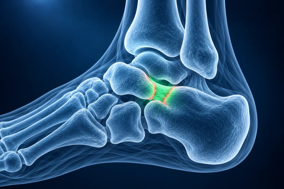
The bridge can be solid bone (osseous), cartilage, or fibrous tissue, and the last two are easy to miss on X-ray. A radiology review published in HSS Journal estimates that coalitions affect up to 13% of people, many of whom never have symptoms, and the American Academy of Orthopaedic Surgeons notes that both feet are affected in about half of cases. This page explains the signs radiologists report and which scan answers which question. It is educational guidance, not a diagnosis.
Why does a tarsal coalition cause a stiff flat foot?
The joints beneath the ankle let the heel roll in and out as you walk on uneven ground. When two of those bones are tied together, that motion is lost, more stress passes to neighboring joints, and the foot tends to settle into a flat position with the heel tilted outward. The muscles on the outer leg may go into protective spasm, which is why older texts call it peroneal spastic flatfoot.
Symptoms often start after a growth spurt, a step up in sport, or a minor sprain that never seems to settle. Some teenagers are first seen for recurrent ankle sprains before anyone checks the hindfoot. A coalition is one of the main causes doctors look for when a flat foot is rigid rather than flexible.
What do “C-sign” and “anteater nose sign” mean on a foot X-ray?
These are the phrases that appear most often on coalition reports, explained in plain words. Coalition terminology can be read alongside the foot X-ray report glossary.
- “Anteater nose sign”: on the side view, the front tip of the heel bone looks long and pointed as it reaches toward the navicular, like an anteater's snout. It suggests a calcaneonavicular coalition.
- “Calcaneonavicular bar on the oblique view”: the 45° oblique X-ray shows the gap between the heel bone and the navicular directly. A bony bar or a narrow, irregular gap with ragged edges points to a coalition.
- “Continuous C-sign”: on the side view, an unbroken C-shaped line runs from the top of the talus down around the shelf of the heel bone that supports it. It suggests a talocalcaneal coalition at the middle facet, which is hard to see directly on plain X-ray.
- “Talar beak”: a small lip of bone on top of the talar head, caused by abnormal stress on the joint in front of the talus. It is a secondary sign of reduced hindfoot motion, not necessarily arthritis.
- “Absent middle facet” or “dysplastic sustentaculum tali”: the normal joint between the talus and the supporting shelf of the heel bone cannot be seen, or the shelf looks broad and misshapen.
- “Non-osseous (fibrous or cartilaginous) coalition with subchondral cysts and marrow edema”: on CT or MRI the joint is narrowed and irregular with small cysts and swelling in the bone on both sides, showing the bridge is not solid bone but is still under strain.
For the views and landmarks behind these signs, see how to read a foot X-ray. If you already have the images, open them in the free X-ray viewer.
Do you need a CT or MRI for a suspected tarsal coalition?
For adults with suspected bony hindfoot disease, ACR guidance starts with radiographs; MRI or CT without contrast is usually appropriate when radiographs do not answer the question.
Standing X-rays come first: top-down, side and 45° oblique views of the foot. A study by Crim and Kjeldsberg found that routine top-down and side views are a valuable screening tool for both main types when the reader looks for the specific signs, but plain X-rays cannot show how far a talocalcaneal bar extends or what tissue it is made of.
CT is the usual next step when surgery is being considered. Thin slices through both feet show exactly which bones are joined, how much of the joint surface is involved, and whether the other foot has a coalition too, which matters because the extent of a talocalcaneal coalition helps decide between removing the bar and fusing the joint. CT studies can be opened in the free CT viewer.
MRI is best for fibrous and cartilage bridges, which show as an irregular, fluid-poor joint with bone marrow edema on both sides, and for other causes of hindfoot pain such as a stress fracture or a tendon problem. It uses no radiation, which matters in children. The foot MRI reading guide explains the sequences reports refer to.
What We Look For on Coalition Imaging
- Bony, cartilaginous or fibrous bridging between the calcaneus and navicular or the talus and calcaneus
- Anteater nose sign, continuous C-sign and talar beak on the standing side view
- Narrowed, irregular joint margins with subchondral cysts or sclerosis at the involved joint
- Bone marrow edema on both sides of a non-osseous coalition on MRI
- The share of the middle facet involved in a talocalcaneal coalition on CT
- Hindfoot valgus, flat-foot alignment and early arthritis in neighboring joints
How is a painful tarsal coalition treated?
Treatment starts without surgery. A break from high-impact activity for a few weeks, sometimes in a walking cast or boot, shoe inserts and anti-inflammatory medicine settle symptoms in many teenagers. A coalition that is found by chance and does not hurt usually needs no treatment at all.
If pain keeps returning, surgeons may remove the bar and fill the gap with fat or muscle so it does not regrow, often correcting the flat foot at the same time. Large talocalcaneal coalitions or a hindfoot that already shows arthritis may be better treated by fusing the joint. Imaging of both feet is part of the planning, because a coalition on one side raises the chance of one on the other.
A tender bump on the inner midfoot in the same age group is more often an accessory navicular, and pain under the heel in an active teenager can come from the growth plate of the heel bone. A clinician examines the foot to tell them apart. Our children’s foot-pain guide compares these childhood symptom patterns.
When should foot pain in a child be seen urgently?
- Fever, a hot red swollen foot or ankle, or a child who refuses to put weight on the foot.
- Pain that wakes the child at night or continues at rest, or unexplained weight loss.
- A foot that becomes stiff or changes shape quickly, or numbness and weakness in the foot.
These patterns are uncommon with a coalition and deserve prompt medical review to rule out infection, inflammatory arthritis or other causes.
Frequently Asked Questions
Can a tarsal coalition be missed on X-ray?
Yes. Fibrous and cartilage coalitions, and many talocalcaneal coalitions at the middle facet, can be hard to see on plain X-rays. Indirect signs such as the C-sign or a talar beak raise suspicion, and CT or MRI is then used to confirm the bridge.
What is the difference between a calcaneonavicular and a talocalcaneal coalition?
A calcaneonavicular coalition joins the front of the heel bone to the navicular and is usually visible on a 45° oblique X-ray. A talocalcaneal coalition joins the talus and heel bone beneath the ankle, most often at the middle facet, and usually needs CT or MRI to measure. They tend to cause symptoms at slightly different ages and may be treated differently.
Does a tarsal coalition always need surgery?
No. Many coalitions never cause pain, and many painful ones settle with rest, a boot or cast and inserts. Surgery is considered when symptoms keep returning despite these measures.
Can adults be diagnosed with a tarsal coalition?
Yes. Some coalitions stay silent through childhood and are found in adults after an ankle sprain, or because of hindfoot arthritis. The imaging signs are the same, although arthritis in nearby joints becomes more important in treatment decisions.
Can AI spot a tarsal coalition on my scan?
X-ray, CT or MRI can show a coalition, while examination checks hindfoot movement and symptoms. AI cannot confirm the diagnosis or decide treatment.
Sources
- Lawrence DA, Rolen MF, Haims AH, Zayour Z, Moukaddam HA. Tarsal Coalitions: Radiographic, CT, and MR Imaging Findings. HSS J. 2014;10(2):153-166.
- Crim JR, Kjeldsberg KM. Radiographic diagnosis of tarsal coalition. AJR Am J Roentgenol. 2004;182(2):323-328.
- Guduri V, Dreyer MA. Talocalcaneal Coalition. In: StatPearls. Treasure Island (FL): StatPearls Publishing.
- American Academy of Orthopaedic Surgeons. Tarsal Coalition. OrthoInfo.
Related Articles
Most plantar fasciitis needs no MRI. Learn when an X-ray, ultrasound or MRI is worth it, what each shows, and what heel pain report phrases mean.
Why a child's foot hurts: Sever's, Köhler, tarsal coalition, accessory navicular and toddler fractures, what X-rays show and the red flags to act on.
What a standing weight-bearing CT of the foot shows that standing X-rays and MRI cannot, who it helps, how its radiation dose compares and where it is offered.
Understand your imaging before your next appointment
MRI, X-ray or CT from a CD, folder or ZIP. A PDF report or photos of the films work too.
Free to open and preview, no account needed. An optional AI report from $10 explains your images in plain language, with questions for your doctor.
Your raw files stay on this device. Only rendered images and minimal context are sent when you request an AI report.
Medical Disclaimer: This page is for informational and educational purposes only. It does not constitute medical advice, diagnosis, or treatment. AI-generated analysis may contain errors. Always consult a qualified healthcare professional for medical decisions. Full Disclaimer