
Metatarsal Stress Fracture: Why the X-ray Looks Normal and What MRI Shows
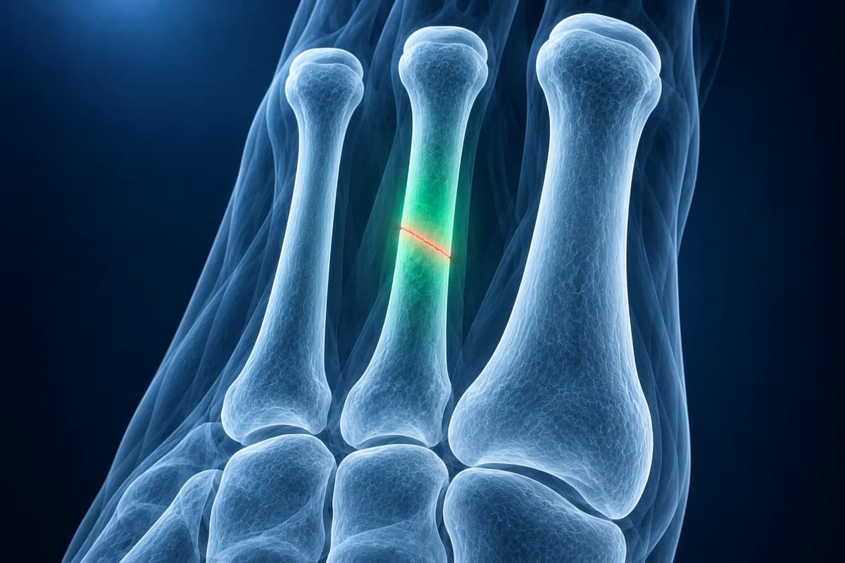
A metatarsal stress fracture is a crack in one of the long forefoot bones caused by repeated loading rather than a single blow. It most often affects the second or third metatarsal. Early X-rays are frequently normal, because the crack only shows once healing bone forms after two to three weeks or more; MRI can show the bone marrow edema of a stress injury within days.
Common Symptoms
- Aching on the top of the forefoot that starts during a run or long walk and eases with rest
- Pain that gradually appears earlier in each session and later lingers after activity stops
- A pinpoint tender spot when pressing on one metatarsal shaft, most often the second or third
- Mild swelling on the top of the foot without a single remembered injury
- A firm bump on the bone after several weeks, which is healing callus
- Pain on hopping or pushing off the forefoot, while walking on the heel feels better
Why do metatarsals crack without an injury?
Bone constantly remodels: small areas of microdamage are removed and rebuilt. When loading rises faster than that repair cycle can keep up with, the microdamage accumulates, the bone first reacts with swelling inside the marrow and around its outer lining, and eventually a crack forms. This is why the classic story is a recent change: a new marathon plan, a switch to minimal shoes, a military training course, a jump in dance rehearsals, or more hours on hard floors.
The second metatarsal is long, thin and held firmly at its base, so it takes much of the push-off load and is the classic site, followed by the third. Factors that make bone less able to cope also matter: low energy intake relative to training, menstrual changes in athletes, low vitamin D, a high-arched or stiff foot, and a short or hypermobile first metatarsal that shifts load to the lesser rays. A clinician may look into bone health when a stress fracture recurs or appears without much extra loading.
When should forefoot pain be seen urgently?
Get same-day care if you suddenly cannot bear weight, if the foot became deformed after a fall or crush, if there is an open wound, or if the toes are numb, pale or cold. Severe, rising pain with a tense, swollen foot after a crush injury can be compartment syndrome and is an emergency. People with diabetes and reduced sensation should also be seen promptly for a warm, swollen forefoot, because a painless fracture can be the first stage of Charcot foot.
Why can my X-ray be normal if I have a stress fracture?
In the first weeks the crack is often a hairline that runs through bone of the same density, so the X-ray beam passes through it with no visible change. What eventually makes it visible is the repair: a fluffy shell of new bone along the outer surface (periosteal reaction) and a hazy band across the shaft. That usually takes two to three weeks or longer to appear, which is why a repeat X-ray is sometimes the simplest next step. A normal first film does not rule out a stress fracture. The full story is in our article on stress fractures that do not show on X-ray.
The ACR Appropriateness Criteria for stress fractures place radiographs first, then MRI when X-rays are normal or unclear and the answer would change management. MRI is the most sensitive test and also shows nearby problems that can feel similar. CT can define a fracture line in bone that is hard to see, and a nuclear bone scan is now used mainly when MRI is not possible.
What does "bone marrow edema in the second metatarsal" mean?
- "Bone marrow edema"
- Extra fluid inside the bone, bright on fluid-sensitive MRI images, usually a stress reaction in a runner with forefoot pain. Ask which metatarsal and which part of it is involved, since the base of the second metatarsal and the fifth metatarsal are higher-risk sites.
- "Periosteal edema"
- Swelling of the thin membrane that wraps the bone, the earliest and mildest stage of a stress injury. Ask how much you need to cut back and for how long.
- "Stress reaction"
- Bone that is overloaded and swollen but has no visible crack yet, the stage where offloading works fastest. Ask what MRI grade the radiologist assigned and how it changes your time away from running.
- "Linear low signal" or "fracture line"
- A dark line through the swollen bone on MRI, meaning an actual stress fracture has formed. Ask whether you need a boot and how long before a follow-up X-ray.
- "Periosteal reaction" or "callus formation"
- X-ray wording for new bone forming around the crack, which usually means the fracture is healing. Ask whether the amount of callus fits the time since your pain started.
For the rest of a foot report, see how to read a foot MRI and foot MRI report phrases explained.
How are stress injuries graded on MRI?
Many radiologists describe severity with a four-grade system first published by Fredericson and colleagues for the shin bone and widely applied to the foot. Grade 1 is swelling of the bone lining only. Grade 2 adds marrow edema on fluid-sensitive images. Grade 3 shows marrow edema on both fluid-sensitive and T1-weighted images. Grade 4 means a visible fracture line. Higher grades generally need longer offloading, but the grade is one input; the bone involved, symptoms and your clinician's examination drive the plan.
What we look for on forefoot stress imaging
- Periosteal and marrow edema around the second, third or fourth metatarsal shaft or neck
- A low-signal line through the edema that upgrades a stress reaction to a fracture
- Periosteal new bone, callus or a sclerotic band on follow-up X-rays
- Involvement of high-risk sites such as the base of the second or fifth metatarsal
- Joint fluid or metatarsal head change that points to a different forefoot diagnosis
Which metatarsal stress fractures are high risk?
Shaft fractures of the second to fourth metatarsals are considered low risk: they usually heal with activity reduction and a stiff-soled shoe or walking boot. Two sites behave differently. The proximal shaft of the fifth metatarsal has a poor blood supply and a known risk of delayed healing, covered on our Jones fracture page. The base of the second metatarsal, common in ballet dancers, sits right at the Lisfranc joint and can be confused with a Lisfranc injury. These usually get closer follow-up and sometimes a period without weight bearing.
How long does a metatarsal stress fracture take to heal?
Most low-risk metatarsal stress fractures settle over about six to eight weeks of protected activity, with a gradual return to running guided by pain-free walking and hopping. Cross-training such as cycling or swimming can usually continue if it is pain free. Returning too early is the most common reason symptoms come back. The metatarsal fracture healing timeline explains what follow-up X-rays show at each stage.
Could it be something other than a stress fracture?
Pain under the ball of the foot rather than on top is more typical of a plantar plate tear or a Morton's neuroma, which causes burning and tingling between the toes. Pain centred on the second metatarsal head in a teenager or young woman can be Freiberg disease. A hot, red, very tender joint can be gout. Imaging helps separate these, which is why MRI is useful when the picture is unclear.
Can I look at my own foot MRI or X-ray?
You can scroll through your scan privately in the free foot MRI viewer or open your films in the free X-ray viewer; files stay on your device. To get the report wording explained in plain language, upload your images for an AI review. It is an explanation, not a diagnosis, and your clinician remains responsible for treatment decisions. Explore ball-of-foot pain for a symptom-based comparison with metatarsal stress pain.
Frequently Asked Questions
Can you walk on a metatarsal stress fracture?
Many people can walk on a low-risk metatarsal stress fracture, which is why it is often ignored for weeks. Walking is usually allowed in a stiff-soled shoe or boot if it is not painful, but running and jumping are paused. High-risk sites such as the proximal fifth metatarsal may need a period without weight bearing, so ask your clinician which applies to you.
Is a stress reaction the same as a stress fracture?
No. A stress reaction is overloaded, swollen bone without a crack, while a stress fracture has a visible fracture line. They are stages of the same process, and a stress reaction that keeps being loaded can progress to a fracture. Both are treated by reducing load; the fracture usually takes longer.
Do I need an MRI for a suspected metatarsal stress fracture?
Not always. If the story and examination are typical, many clinicians treat a presumed low-risk stress fracture and repeat the X-ray after a few weeks. MRI is most useful when an early answer matters, such as for athletes in season, high-risk locations, or pain that does not fit a stress fracture.
How long does bone marrow edema take to go away on MRI?
Marrow edema often fades more slowly than the pain, sometimes lingering for months after you are back to normal activity. For that reason, return to sport is usually guided by symptoms, examination and X-ray healing rather than by a repeat MRI.
Sources
- Expert Panel on Musculoskeletal Imaging; Morrison WB, Deely D, Fox MG, et al. ACR Appropriateness Criteria Stress (Fatigue-Insufficiency) Fracture Including Sacrum Excluding Other Vertebrae: 2024 Update. J Am Coll Radiol. 2024;21(11S):S490-S503.
- Fredericson M, Bergman AG, Hoffman KL, Dillingham MS. Tibial stress reaction in runners. Correlation of clinical symptoms and scintigraphy with a new magnetic resonance imaging grading system. Am J Sports Med. 1995;23(4):472-481.
- Bergman R, Kaiser K. Stress Reaction and Fractures. StatPearls. Treasure Island (FL): StatPearls Publishing.
- American Academy of Orthopaedic Surgeons. Stress Fractures of the Foot and Ankle. OrthoInfo.
Related Articles
Why Lisfranc midfoot injuries are missed on first X-rays, the alignment lines and fleck sign to check, and when standing X-rays, CT or MRI are needed.
How long a broken metatarsal takes to heal week by week, why the fracture line lingers on X-ray, which fractures heal slowly, and what follow-up reports mean.
Why a foot stress fracture can hide on a normal X-ray, what MRI, CT and repeat X-rays show instead, and which foot stress fractures are high risk.
Understand your imaging before your next appointment
MRI, X-ray or CT from a CD, folder or ZIP. A PDF report or photos of the films work too.
Free to open and preview, no account needed. An optional AI report from $10 explains your images in plain language, with questions for your doctor.
Your raw files stay on this device. Only rendered images and minimal context are sent when you request an AI report.
Medical Disclaimer: This page is for informational and educational purposes only. It does not constitute medical advice, diagnosis, or treatment. AI-generated analysis may contain errors. Always consult a qualified healthcare professional for medical decisions. Full Disclaimer