
Foot MRI Report Phrases Explained in Plain Language
What bone marrow edema, plantar fascia thickening, Lisfranc ligament sprain, plantar plate attenuation and sesamoid edema mean on your foot MRI report.
Key Takeaways
- Read the Impression first: it ranks the findings the body of the report lists
- Bone marrow edema in a metatarsal shaft usually means a stress reaction; a fracture line means a stress fracture
- On ultrasound, a plantar fascia thicker than about 4 mm at the heel is a well-established sign of plantar fasciitis
- Attenuation of a plantar plate is thinning, often before a full tear, most commonly at the second toe
- Infection words such as osteomyelitis, abscess or sinus tract need same-day medical contact
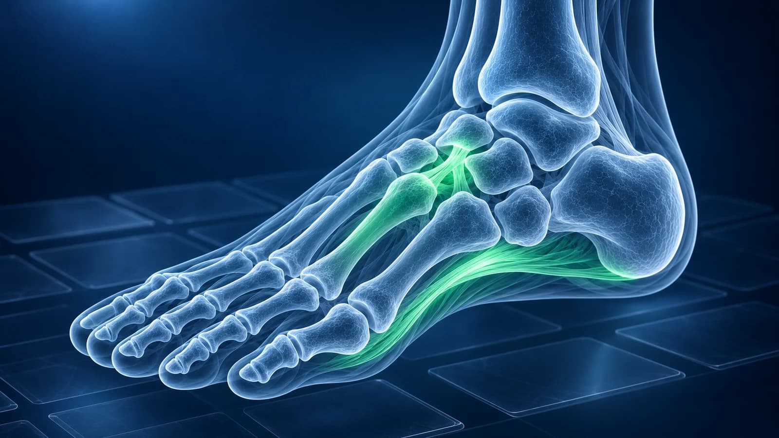
Most foot MRI report phrases describe one of three things: water where it should not be (“edema”), a structure that is thicker or thinner than normal (“thickening”, “attenuation”), or a gap in a ligament or bone (“tear”, “disruption”, “fracture line”). On ultrasound, for example, a plantar fascia thicker than about 4 mm at the heel is a well-established sign of plantar fasciitis. The phrase only becomes a diagnosis when your clinician matches it to where it hurts.
This page takes the phrases foot MRI reports print most often, one by one, and says what each one usually means, which condition it tends to belong to and what to ask the person who ordered the scan. It explains wording; it does not review your images and is not a diagnosis.
Where in the foot do the common report phrases point?
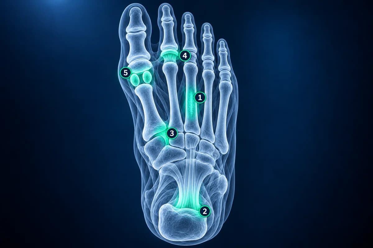
Reports are usually written from the top of the scan down, not in order of importance. The sentence that matters most is often in the “Impression” or “Conclusion” at the end, where the radiologist ranks the findings. Read that part first, then find each finding in the body of the report using the numbered map above.
What does “bone marrow edema in the second metatarsal” mean?
It means the inside of the bone holds more water than normal, which shows up bright on the fluid-sensitive (STIR) images. In a runner, walker or someone who recently changed shoes or training, edema along the shaft of the second or third metatarsal usually means a “stress reaction”: the bone is being loaded faster than it can repair. If the report adds “a low-signal fracture line”, the reaction has progressed to a metatarsal stress fracture. The same pattern in the navicular, on the top of the midfoot, is handled more cautiously; see navicular stress fracture.
Ask your clinician: is there a fracture line or only edema, how much should I reduce loading and for how long, and is a follow-up image needed? An X-ray taken in the first weeks is often normal, which is explained in why a stress fracture may not show on X-ray.
What does “thickening of the plantar fascia” mean?
The plantar fascia is the tough band under the arch that starts at the heel bone. On MRI a healthy band is thin and uniformly dark; in plantar fasciitis it is thickened near the heel (on ultrasound, more than about 4 mm is the usual cut-off), and the report may add “perifascial edema” (fluid around it) or “calcaneal enthesis edema” (swelling in the heel bone where it attaches). A firm, rounded lump further forward in the band, away from the heel, is described differently, as a “fibroma”; see plantar fibroma.
Ask your clinician: is there a partial tear as well as thickening, and does the finding change the plan from stretching, footwear and load management? Most heel pain is managed without a scan at all.
What does “Lisfranc ligament sprain” or “disruption” mean?
The Lisfranc ligament is a short, strong band that ties the base of the second metatarsal to the medial cuneiform. It holds the arch of the midfoot together. “Sprain” or “partial tear” means some fibres are stretched or torn but the band is in continuity; “disruption” or “complete tear” means it no longer connects the two bones. The report may also note edema in the cuneiforms or metatarsal bases and whether the bones are still lined up. These details decide whether a Lisfranc injury is stable.
Ask your clinician: is the joint stable, is a standing X-ray of both feet needed to compare the gap, and does this need a foot and ankle surgeon’s opinion? A disruption with any widening is usually treated very differently from a sprain.
What does “plantar plate attenuation” mean?
The plantar plate is the thick pad of fibrous tissue on the underside of each lesser toe joint. “Attenuated” means it is thinned or frayed, usually near its attachment on the toe bone, without a clear full-thickness gap. It is often the early stage of a plantar plate tear, most often at the second toe, and the report may add “pericapsular fibrosis” or note that the toe is starting to drift. Pain under that joint without a plate finding is often grouped under metatarsalgia.
Ask your clinician: is it attenuation or a tear, is the toe unstable when examined, and would taping, a stiffer shoe or a metatarsal pad be tried first?
What is the difference between “intermetatarsal bursitis” and a neuroma?
Both sit in the web space between two metatarsal heads, usually the third, and both can cause burning or numb toes. A bursa is a small fluid-filled sac, so “intermetatarsal bursitis” is bright on fluid-sensitive images. A neuroma is a thickened nerve with scar tissue around it, so it is dark, teardrop-shaped and best seen on T1 short-axis slices. Reports often describe both together, as the bursa commonly sits just above the nerve in Morton’s neuroma.
Ask your clinician: what size is the neuroma, does it match the web space where my toes go numb, and would an injection be guided by ultrasound? Pooled studies find ultrasound and MRI similarly accurate for this lump, as covered in Morton’s neuroma ultrasound vs MRI.
What does “sesamoid edema” mean?
The sesamoids are two pea-sized bones inside the tendon under the big toe joint. Edema in one of them, usually the inner (medial) one, fits overload and sesamoiditis. Words such as “fracture” or “fragmentation” point to a broken sesamoid or one that has lost its blood supply (avascular necrosis) instead. Many people are born with a sesamoid in two parts (“bipartite”), which can be confused with a fracture. After a forced bend of the big toe, edema with a torn capsule beneath the joint suggests turf toe.
Ask your clinician: is this edema alone, a fracture or a bipartite sesamoid, and how long should I offload the ball of the foot?
Why does a foot MRI report mention “Kager’s fat pad”?
Kager’s fat pad is the triangle of fat in front of the Achilles tendon, just above the heel bone. It appears on sagittal images of a hindfoot study. “Edema in Kager’s fat pad” is a sign of irritation next to the Achilles tendon or the bursa behind the heel bone, so it belongs to the ankle more than to the foot. Our ankle site covers the Achilles tendon tear and the wider tendon picture. Ask your clinician whether the Achilles tendon itself is thickened or torn.
When is an MRI ordered for foot pain in the first place?
The American College of Radiology recommends plain X-rays as the first test for chronic foot pain and after most foot injuries. MRI is usually added when the X-rays are normal or unclear and a stress fracture, a ligament or plantar plate injury, a neuroma or an infection is still suspected. Ultrasound is an alternative for superficial structures such as the plantar fascia, a neuroma or a plantar plate. CT is preferred when the fine shape of a fracture or joint matters. MRI cannot show how a joint behaves when you stand, which is why a standing X-ray may still be requested afterwards. The trade-offs are laid out in foot MRI vs X-ray vs ultrasound.
Which foot MRI findings need prompt care?
Contact a clinician the same day if your report mentions “osteomyelitis”, an “abscess”, “septic arthritis” or a “sinus tract”, especially if you have diabetes, a wound on the foot, fever, or a foot that is hot, red and swollen. The same applies to a Lisfranc disruption with displaced bones. Do not wait for a routine follow-up to discuss these phrases.
How can I find the finding on my own images?
Open the DICOM files from your disc in our free MRI viewer, which runs in your browser and keeps the files on your device. Choose a STIR or fat-suppressed series and scroll to the bone or space the report names; the bright area should be easy to spot. For the sequences and planes themselves, start with how to read a foot MRI.
Frequently Asked Questions
Is “stress reaction” the same as a stress fracture?
No. A stress reaction is edema in the bone without a visible fracture line; a stress fracture shows a line through the bone. They sit on one spectrum, and reducing load early can stop a reaction from becoming a fracture.
What does “nonspecific” mean on my foot MRI report?
It means the finding can have several causes and the image alone cannot choose between them. Your clinician uses your symptoms, the examination and sometimes blood tests or a follow-up scan to narrow it down.
What does “small joint effusion” at a toe joint mean?
A little fluid in a toe joint is common and often harmless. It matters more when it is large, sits in a joint that hurts, or appears with edema in both bones of the joint, which can point to arthritis, gout or, rarely, infection.
Why does my report list findings I have no pain from?
Radiologists describe everything they see, including small spurs, bursae and cysts that many people without symptoms also have. The finding that explains your pain is usually the one that matches the spot that hurts.
Can AI explain my foot MRI report phrases?
AI can translate the terms and show which structure each one refers to, which helps you prepare questions. It cannot examine your foot or confirm a diagnosis; that remains with your clinician.
Sources
- Expert Panel on Musculoskeletal Imaging; Said N, Fox MG, Nacey NC, et al. ACR Appropriateness Criteria Chronic Foot Pain: Update 2025. J Am Coll Radiol. 2026;23(4):681-699.
- Expert Panel on Musculoskeletal Imaging; Gorbachova T, Chang EY, Ha AS, et al. ACR Appropriateness Criteria Acute Trauma to the Foot. J Am Coll Radiol. 2020;17(5S):S2-S11.
- McMillan AM, Landorf KB, Barrett JT, Menz HB, Bird AR. Diagnostic imaging for chronic plantar heel pain: a systematic review and meta-analysis. J Foot Ankle Res. 2009;2:32.
- Albright RH, Brooks BM, Chingre M, et al. Diagnostic accuracy of magnetic resonance imaging (MRI) versus dynamic ultrasound for plantar plate injuries: a systematic review and meta-analysis. Eur J Radiol. 2022;152:110315.
- Bignotti B, Signori A, Sormani MP, et al. Ultrasound versus magnetic resonance imaging for Morton neuroma: systematic review and meta-analysis. Eur Radiol. 2015;25(8):2254-2262.
- Sims AL, Kurup HV. Painful sesamoid of the great toe. World J Orthop. 2014;5(2):146-150.
Related Articles
A plain-language guide to your foot MRI: how T1, fluid-sensitive and STIR images differ, which plane shows what, and what bone marrow edema, plantar plate tear and neuroma mean in your report.
X-ray, MRI, ultrasound or CT for foot pain? What each test shows and misses, which one usually comes first, and how the choice changes for stress fractures, neuromas, plantar fasciitis, Lisfranc injuries and the diabetic foot.
Why a foot stress fracture can hide on a normal X-ray, what MRI, CT and repeat X-rays show instead, and which foot stress fractures are high risk.
Related Conditions
Understand your imaging before your next appointment
MRI, X-ray or CT from a CD, folder or ZIP. A PDF report or photos of the films work too.
Free to open and preview, no account needed. An optional AI report from $10 explains your images in plain language, with questions for your doctor.
Your raw files stay on this device. Only rendered images and minimal context are sent when you request an AI report.
Medical Disclaimer: This page is for informational and educational purposes only. It does not constitute medical advice, diagnosis, or treatment. AI-generated analysis may contain errors. Always consult a qualified healthcare professional for medical decisions. Full Disclaimer