
Morton's Neuroma: Ultrasound vs MRI
Which scan finds a Morton's neuroma better, what each one shows and misses, how reports describe it, and when MRI is worth the extra cost.
Key Takeaways
- Ultrasound and MRI have similar sensitivity for Morton's neuroma, around 90%
- A standing X-ray usually comes first to exclude bone causes of forefoot pain
- Ultrasound is quick, dynamic and can guide an injection, but depends on the operator
- MRI is best when the cause is unclear, because it also shows bones, joints and plantar plates
- A reported neuroma must match your symptoms; small ones are common in people without pain
Ultrasound and MRI detect a Morton's neuroma about equally well: a meta-analysis of surgically confirmed cases found a sensitivity of about 91% for ultrasound and 90% for MRI. Ultrasound is usually chosen first because it is cheaper, quick and can be done while you squeeze the forefoot; MRI is used when the diagnosis is unclear or other causes of ball-of-foot pain need ruling out.
In other words, the choice is less about which machine is better and more about which question is being asked. If the story and examination already point strongly to a neuroma, ultrasound confirms it and can guide an injection in the same visit. If the pain could equally be a joint, a ligament or a bone problem, MRI looks at all of them at once. The sections below compare the two tests in detail; they are general education, not a substitute for the clinician examining your foot.
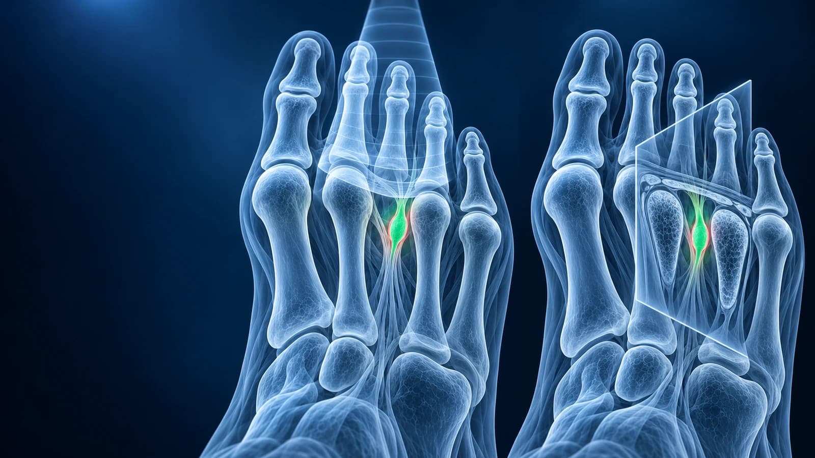
What exactly is the scan looking for?
A Morton's neuroma is not a true tumour. It is a thickening of a common plantar digital nerve, with scar-like fibrous tissue around it, where the nerve passes beneath the deep transverse metatarsal ligament between two metatarsal heads. It most often sits in the third web space, between the third and fourth toes, and less often in the second. The typical story is burning or electric pain in the ball of the foot that shoots into two neighbouring toes, a feeling of walking on a pebble or a folded sock, and relief when tight shoes come off.
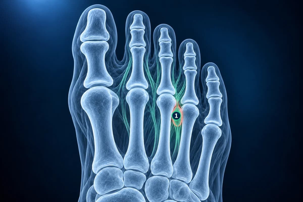
The lesion is small, usually a few millimetres to around a centimetre, and it lies in a tight space crowded with other structures that can cause similar pain: the joint capsules and plantar plates of the toes, a fluid-filled intermetatarsal bursa, and the metatarsal bones themselves. A useful scan has to show the neuroma and, just as importantly, show that its neighbours are normal.
Which test comes first for suspected Morton's neuroma?
The diagnosis is often clinical, and many people are treated without any scan. When imaging is needed, the ACR Appropriateness Criteria for chronic foot pain start with a standing X-ray. It cannot show the nerve, but it can show what else might be causing the pain: a metatarsal stress fracture, a flattened metatarsal head from Freiberg disease, arthritis, or toes drifting apart. After that, ultrasound and MRI are both considered appropriate next steps, and local availability and expertise often decide between them.
What does ultrasound show in Morton's neuroma?
On ultrasound a neuroma appears as a darker, oval, poorly compressible nodule in the web space, often scanned from the sole with the probe aligned along the toes. Because the examination is live, the sonographer can press the metatarsal heads together and watch the nodule pop downward, the sonographic version of the Mulder click felt in clinic. Pressing directly on the lesion and reproducing your exact pain adds confidence that the finding matters.
Ultrasound has practical advantages: it is quick, involves no radiation or enclosed scanner, compares both feet and every web space in minutes, and can guide a needle precisely if an injection is planned. Its weakness is that it depends on the person holding the probe. Very small neuromas, a foot with lots of scar tissue after previous surgery, or a lesion obscured by a bursa can be missed or overcalled, and ultrasound shows little of the bone marrow or deeper joint structures.
What does MRI show in Morton's neuroma?
On MRI the neuroma is best seen on short-axis images that slice across the forefoot, where it looks like a teardrop of tissue hanging down between two metatarsal heads, below the transverse ligament. Because it is mostly fibrous, it is typically dark on both T1 and T2 images, which helps separate it from a bright, fluid-filled bursa. Some centres scan the patient lying face down so the forefoot is not squashed, and some give contrast, which can make the lesion stand out.
The real strength of MRI is the wider view. In the same study it can show bone marrow edema from a stress reaction, a plantar plate tear under the second toe, inflammation of a toe joint, or a different soft-tissue mass. The trade-offs are cost, time in the scanner and a static picture that cannot reproduce the click or your pain. The guide to reading a foot MRI explains the planes and sequences, and foot MRI cost covers what drives the price.
Ultrasound or MRI: which is better for you?
- Accuracy for the neuroma itself: comparable in pooled studies, with MRI reporting slightly fewer false positives in the small number of studies available
- Classic symptoms in one web space: ultrasound is usually enough and is often the more cost-effective choice
- Unclear pain, two web spaces involved, or pain under the second toe: MRI shows the alternative causes as well
- Planning an injection: ultrasound can confirm the lesion and guide the needle at the same visit
- Before surgery or after failed surgery: MRI is often preferred to map the lesion and look for a stump neuroma or another cause
A broader comparison of all three foot tests is in foot MRI vs X-ray vs ultrasound.
What do Morton's neuroma reports say?
- "Hypoechoic ovoid lesion in the third intermetatarsal space measuring 7 × 4 mm" — the ultrasound description of a neuroma with its size
- "Positive sonographic Mulder sign" — the lesion moved out of the web space when the forefoot was squeezed, which supports the diagnosis
- "Teardrop-shaped T1 and T2 hypointense lesion plantar to the deep transverse metatarsal ligament" — the MRI wording for the same thing
- "Intermetatarsal bursal fluid" — a small fluid sac in the web space, which can accompany a neuroma or cause similar pain on its own
- "No bone marrow edema; plantar plates intact" — the bones and the toe joint supports look normal, which makes a neuroma a more likely cause of the pain
One caution applies to both tests: small neuromas are also found in people who have no foot pain at all. A reported lesion therefore needs to match where you hurt and what the examination finds. Larger lesions and those that reproduce your pain are more likely to be the real cause.
If you already have an MRI on disc, you can open the study privately in the free foot MRI viewer, which runs in your browser without uploading the files.
When is ball-of-foot pain not a neuroma?
Most forefoot pain is not urgent, but some patterns should be seen promptly rather than waiting for a routine scan: swelling, warmth and redness across the forefoot, especially in someone with diabetes or reduced feeling; pain that started suddenly with an inability to bear weight; numbness spreading beyond two toes or into the whole foot; a lump that is growing; or fever. These point toward infection, fracture, nerve problems further up the leg or a different mass, and they change which test is needed.
Frequently Asked Questions
Can an X-ray show a Morton's neuroma?
No. A neuroma is soft tissue and is invisible on X-ray. The X-ray is still useful because it can show a stress fracture, Freiberg disease, arthritis or toe malalignment that might explain the same pain.
My ultrasound was normal but the pain continues. Should I have an MRI?
It is a reasonable question to ask your clinician. A normal ultrasound makes a large neuroma unlikely but does not rule out a small one or other causes such as a plantar plate tear or stress reaction. MRI is often the next step when symptoms persist and the diagnosis is still uncertain.
Does the size of the neuroma matter?
Size helps but does not decide everything. Larger neuromas are more likely to cause symptoms, and size is sometimes considered when planning treatment, but pain level does not track size closely and small lesions can be incidental. The match between the scan, your symptoms and the examination matters most.
Is contrast needed for a Morton's neuroma MRI?
Not always. Many centres diagnose neuromas on non-contrast images, while others add contrast because it can make the lesion easier to see. The radiology team decides based on the question on the request form and your kidney function and allergy history.
Can AI read my forefoot MRI?
An AI explanation can describe what is visible on your images and translate report wording into plain language, which may help you prepare questions. It is not a diagnosis, and it does not replace the radiologist's report or the clinician who examines your foot.
Sources
- Bignotti B, Signori A, Sormani MP, et al. Ultrasound versus magnetic resonance imaging for Morton neuroma: systematic review and meta-analysis. Eur Radiol. 2015;25(8):2254-2262.
- Dixon M, Munir U, Tafti D. Morton Neuroma. StatPearls. Treasure Island (FL): StatPearls Publishing.
- Expert Panel on Musculoskeletal Imaging. ACR Appropriateness Criteria Chronic Foot Pain: Update 2025. J Am Coll Radiol. 2026;23(4):681-699.
Related Articles
X-ray, MRI, ultrasound or CT for foot pain? What each test shows and misses, which one usually comes first, and how the choice changes for stress fractures, neuromas, plantar fasciitis, Lisfranc injuries and the diabetic foot.
A plain-language guide to your foot MRI: how T1, fluid-sensitive and STIR images differ, which plane shows what, and what bone marrow edema, plantar plate tear and neuroma mean in your report.
What a foot MRI typically costs, why forefoot, midfoot and contrast scans are billed differently, how insurance applies, and how to lower your bill.
Related Conditions
Understand your imaging before your next appointment
MRI, X-ray or CT from a CD, folder or ZIP. A PDF report or photos of the films work too.
Free to open and preview, no account needed. An optional AI report from $10 explains your images in plain language, with questions for your doctor.
Your raw files stay on this device. Only rendered images and minimal context are sent when you request an AI report.
Medical Disclaimer: This page is for informational and educational purposes only. It does not constitute medical advice, diagnosis, or treatment. AI-generated analysis may contain errors. Always consult a qualified healthcare professional for medical decisions. Full Disclaimer