
How to Read a Foot MRI: Sequences, Planes and Report Phrases
A plain-language guide to your foot MRI: how T1, fluid-sensitive and STIR images differ, which plane shows what, and what bone marrow edema, plantar plate tear and neuroma mean in your report.
Key Takeaways
- T1 shows anatomy and marrow; STIR or fat-suppressed images make swelling and fluid bright
- Short-axis slices show the metatarsal heads in a row, ideal for neuromas and stress fractures
- Bone marrow edema is a sign whose shape and location point to stress, overload, injury or infection
- In a diabetic foot, dark marrow on T1 next to an ulcer is the key MRI sign of osteomyelitis
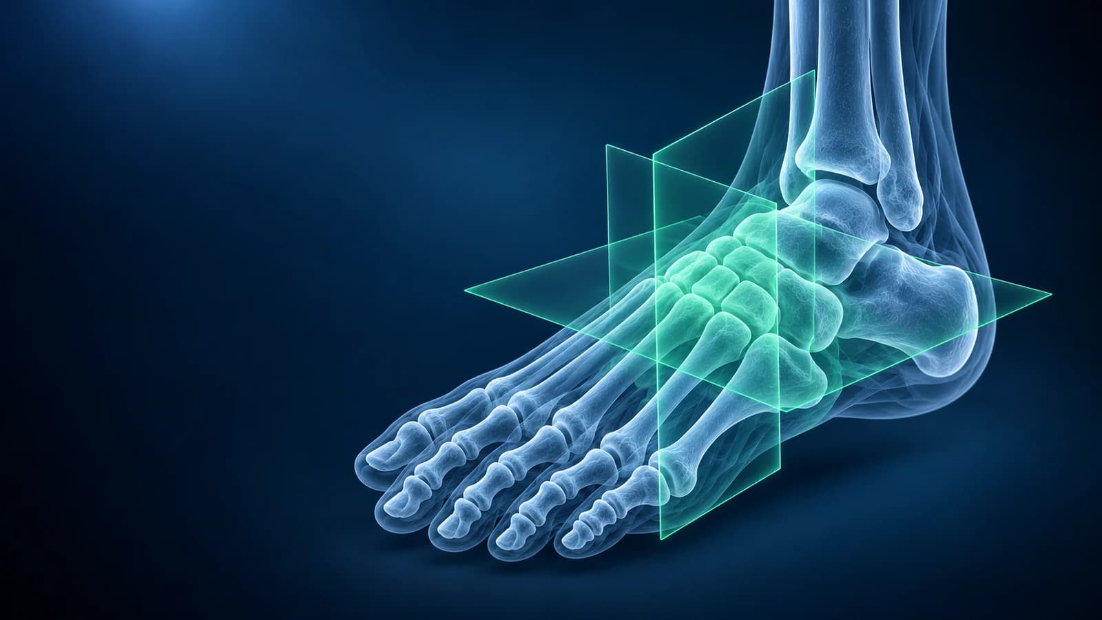
To read a foot MRI, compare two kinds of pictures: T1 images, where healthy bone marrow looks bright, and fluid-sensitive images (STIR or fat-suppressed T2), where water, swelling and inflammation look bright. A spot that is bright on the fluid-sensitive images inside a bone is “bone marrow edema”, the single most common phrase in foot MRI reports, and the location of that spot usually explains the pain.
Foot MRI is ordered when an X-ray has not explained the problem: a suspected stress fracture, a lump between the toes, pain under the ball of the foot, a stubborn heel, or a possible bone infection. This guide covers how the scan is built, how to orient yourself and what the usual report phrases mean. It is educational, not a diagnosis.
Which MRI sequences matter in a foot scan?
- T1-weighted: the anatomy map. Fat and healthy marrow are bright, so a dark patch replacing the marrow is important, especially when infection is a question.
- STIR or fat-suppressed T2 / PD: the problem finder. Fat is turned dark, so edema, fluid in a joint or bursa, and torn tissue light up.
- Proton density without fat suppression: fine detail of tendons, ligaments and the plantar plate, which should look uniformly dark.
- Post-contrast T1 (only sometimes): gadolinium highlights inflamed tissue, abscesses and a neuroma when the plain images are not conclusive.
What are the short-axis and long-axis planes on a foot MRI?
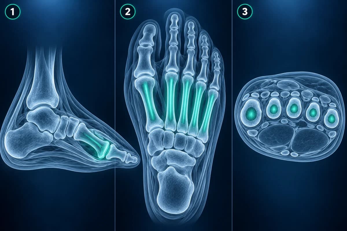
Foot MRI uses its own names for the planes, which confuses many people who have read about knee or spine scans. Short-axis slices cut across the foot like slicing a loaf of bread, so the five metatarsal heads appear as a row of circles; this is where a neuroma between two heads or a stress fracture in a shaft is easiest to see. Long-axis slices run flat along the sole from heel to toes and show the toe joints and the Lisfranc joints in a single view. Sagittal slices look at the foot from the side and are best for the plantar fascia, the arch and the plantar plates under each toe joint.
The field of view also matters. A dedicated forefoot study gives sharp detail of the toes and metatarsal heads but may cut off the heel, and a hindfoot study often belongs to an ankle protocol. If the part that hurts is not on the images, say so; a second targeted scan may be more useful than over-reading the first.
What does “bone marrow edema” in the foot mean?
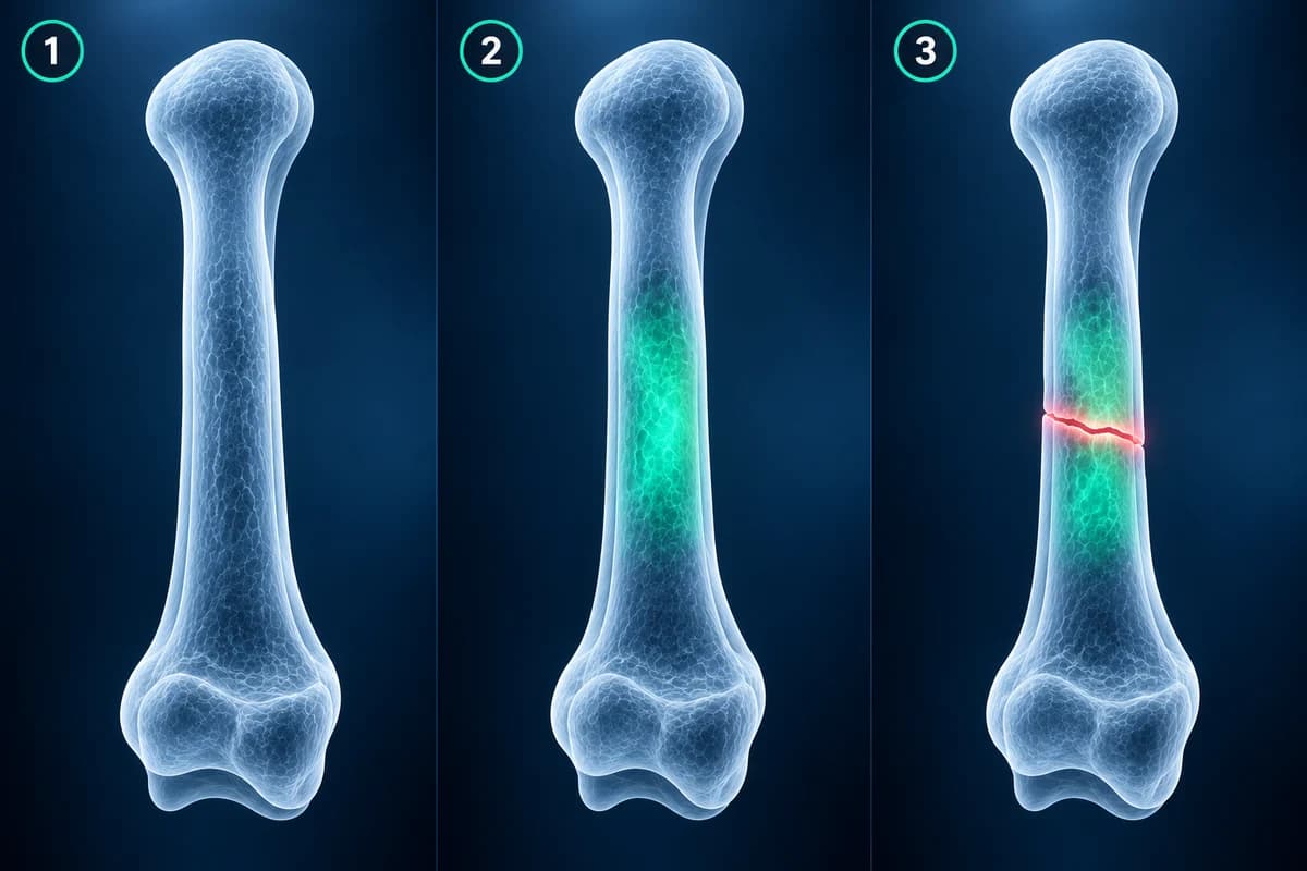
It means extra water inside the bone, seen as brightness on fluid-sensitive images. It is a sign, not a diagnosis, and its shape tells the story. A band of edema across a metatarsal shaft, with or without a thin dark line, fits a stress reaction or a metatarsal stress fracture. Edema in both bones on either side of a joint fits arthritis or overload. Edema in a sesamoid under the big toe points to sesamoiditis or a sesamoid fracture. Edema around the cuneiforms and second metatarsal base after a twist can accompany a Lisfranc injury, where the report should also comment on the Lisfranc ligament itself.
What does a foot MRI show between and under the toes?
The forefoot is where foot MRI does much of its work. An “intermetatarsal neuroma”, most often in the third web space, is a small dark teardrop between two metatarsal heads on short-axis images; reports usually give its size and may mention an “intermetatarsal bursa” beside it. Read more about Morton’s neuroma and why many clinicians start with ultrasound before MRI for a neuroma.
Under each lesser toe joint, the plantar plate is a thick, dark fibrous pad. A bright cleft through it near its attachment on the toe bone is a plantar plate tear, most often at the second toe, and the report may add that the toe is drifting upward or sideways. Pooled studies show dynamic ultrasound is more sensitive for this injury, while MRI is more specific and better for grading it.
What does a foot MRI show in the heel and arch?
On sagittal images the plantar fascia is a thin dark band running from the heel bone toward the toes. In plantar fasciitis the report describes “thickening of the proximal plantar fascia with perifascial edema”, sometimes with edema in the heel bone where it attaches; a partial tear appears as a bright gap within the band. Most heel pain is diagnosed without a scan, which is covered in do you need an MRI for plantar fasciitis. In the midfoot, MRI can show a fibrous or cartilaginous bar between two tarsal bones in tarsal coalition, and edema across the junction of an accessory navicular when it has become painful. Tendons that cross from the ankle into the foot, such as the posterior tibial tendon, are often reported on the same images.
How does MRI tell bone infection from other swelling in a diabetic foot?
Radiologists look at the T1 images. In osteomyelitis the normally bright marrow is replaced by a confluent dark area, often next to an ulcer or a “sinus tract” leading down from the skin. Edema that stays bright on T1 is more likely reactive swelling or a stress change. Reports may also mention an “abscess” (a walled-off fluid collection) or “septic arthritis” of a toe joint. Telling infection from Charcot foot can be difficult, and the location helps: infection usually spreads from a skin wound under a pressure point, while Charcot changes centre on the midfoot joints. See diabetic foot osteomyelitis for the wider picture.
A hot, red, swollen foot in someone with diabetes, a wound with pus or a smell, fever, or a toe turning dark are reasons for same-day medical care, before any MRI is booked or read.
Can I view my own foot MRI slices?
Yes. Load the DICOM files from your disc into our free MRI viewer, which opens them in your browser without sending the original files anywhere. Pick a fluid-sensitive series (labelled STIR, T2 FS or PD FS), find the short-axis stack and scroll to the area the report names. If you are still weighing whether to have the scan, our page on foot MRI cost explains what drives the price.
Frequently Asked Questions
Is bone marrow edema in the foot serious?
It depends on the cause. Edema from overload or a stress reaction often settles with reduced loading over weeks, while edema with a fracture line, a collapsed bone or a nearby ulcer needs a specific treatment plan. Your clinician interprets it together with your symptoms.
Why does my foot MRI not show my heel?
Many foot MRIs are planned as forefoot studies to get sharp detail of the toes and metatarsals. The heel may be only partly included or not at all. If your pain is in the heel, ask whether a hindfoot or ankle protocol was needed.
Do I need contrast for a foot MRI?
Usually not. Contrast is mainly added when infection, an abscess, a mass or inflammatory arthritis is being assessed, or when the plain images leave a question open. The radiologist decides based on the referral.
Can a foot MRI miss a fracture?
MRI is very sensitive to the edema that surrounds fractures, including early stress fractures that X-ray misses. Fine cortical detail and fracture displacement are sometimes clearer on CT, which is why complex midfoot or heel fractures may get both.
Can AI explain my foot MRI report?
AI can translate report terms and point to the structures they describe, which helps you prepare for an appointment. It is not a radiologist and does not replace the clinician who knows your history and examined your foot.
Sources
- Expert Panel on Musculoskeletal Imaging; Said N, Fox MG, Nacey NC, et al. ACR Appropriateness Criteria Chronic Foot Pain: Update 2025. J Am Coll Radiol. 2026;23(4):681-699.
- Expert Panel on Musculoskeletal Imaging; Baker JC, Northrup BE, Ahlawat S, et al. ACR Appropriateness Criteria Suspected Osteomyelitis of the Foot in Patients With Diabetes Mellitus: 2025 Update. J Am Coll Radiol. 2025;22(11S):S713-S737.
- Albright RH, Brooks BM, Chingre M, et al. Diagnostic accuracy of magnetic resonance imaging (MRI) versus dynamic ultrasound for plantar plate injuries: a systematic review and meta-analysis. Eur J Radiol. 2022;152:110315.
- Bignotti B, Signori A, Sormani MP, et al. Ultrasound versus magnetic resonance imaging for Morton neuroma: systematic review and meta-analysis. Eur Radiol. 2015;25(8):2254-2262.
Related Articles
X-ray, MRI, ultrasound or CT for foot pain? What each test shows and misses, which one usually comes first, and how the choice changes for stress fractures, neuromas, plantar fasciitis, Lisfranc injuries and the diabetic foot.
Why a foot stress fracture can hide on a normal X-ray, what MRI, CT and repeat X-rays show instead, and which foot stress fractures are high risk.
What a foot MRI typically costs, why forefoot, midfoot and contrast scans are billed differently, how insurance applies, and how to lower your bill.
Related Conditions
Understand your imaging before your next appointment
MRI, X-ray or CT from a CD, folder or ZIP. A PDF report or photos of the films work too.
Free to open and preview, no account needed. An optional AI report from $10 explains your images in plain language, with questions for your doctor.
Your raw files stay on this device. Only rendered images and minimal context are sent when you request an AI report.
Medical Disclaimer: This page is for informational and educational purposes only. It does not constitute medical advice, diagnosis, or treatment. AI-generated analysis may contain errors. Always consult a qualified healthcare professional for medical decisions. Full Disclaimer