
Sesamoiditis: Telling a Bipartite Sesamoid From a Fracture
Sesamoiditis is pain and irritation of the two small sesamoid bones under the big toe joint. On imaging, the key question is whether a split sesamoid is a harmless bipartite variant, with smooth rounded edges, or a fracture, with sharp jagged edges. X-rays, including a special sesamoid view, come first; MRI shows bone marrow edema, stress injury or loss of blood supply when X-rays are unclear.
Common Symptoms
- Pain on a small spot under the ball of the foot, directly beneath the big toe joint
- Pain that builds with push-off, jumping, dancing on the forefoot or wearing heels
- Pain when the big toe is bent upward, stretching the tendons that hold the sesamoids
- Mild swelling or bruising under the big toe joint, sometimes with a slow, gradual onset
- Walking on the outer edge of the foot to avoid loading the ball of the foot
- A sudden sharp pain after a landing or a forced upward bend of the big toe, if a sesamoid has broken
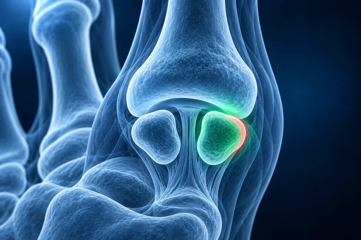
The sesamoids act like a kneecap for the big toe: they lift the tendons that bend it and take much of the load at push-off. Sesamoiditis is used loosely for any painful sesamoid, so the report words matter. This page explains them, without replacing an examination.
My X-ray says “bipartite sesamoid”. Is it broken?
Usually not. Some people's sesamoids develop in two (bipartite) or more pieces and never join. This is a normal variant, most often seen in the inner (tibial) sesamoid and frequently present in both feet. It looks split on an X-ray, which is why it is so often confused with a fracture. Radiologists separate the two by the shape of the gap and the edges:
- Bipartite sesamoid: smooth, rounded pieces, each with its own crisp white outline (cortex), and the pieces together are usually larger than the other sesamoid.
- Fractured sesamoid: a sharp, irregular or jagged line with no outline along the break, pieces that would fit back together like a puzzle, and an overall size similar to the other sesamoid.
- Separation that increases on follow-up films, or a gap that widens on a forced upward-bend view, suggests an injury through the variant rather than a quiet variant.
An X-ray of the other foot is sometimes taken for comparison. If the other side also shows a bipartite sesamoid, the painful side is more likely a variant too, but because the variant is not always present on both sides, a normal other foot does not prove a fracture.
What other phrases appear on a sesamoid report?
- “Bone marrow edema in the tibial sesamoid”: on MRI the bone looks bright on fluid-sensitive images, a sign of stress, bruising or early fracture. It is the classic MRI finding in an overloaded sesamoid.
- “Sesamoid stress fracture”: a thin line through the bone, often with surrounding edema, caused by repeated loading rather than a single injury.
- “Fragmentation” or “sclerosis” of the sesamoid: the bone looks broken into pieces or unusually dense, which can point to osteonecrosis, a loss of blood supply to the bone.
- “Sesamoid-metatarsal arthrosis”: wear of the small joint between the sesamoid and the underside of the first metatarsal head, with narrowing and spurs.
- “Flexor hallucis brevis tendinosis” or “peritendinous edema”: irritation in the tendons that wrap the sesamoids, which can cause similar pain with a normal bone.
What We Look For on Sesamoid Imaging
- Edges of any split: rounded and corticated or sharp and jagged
- Combined size of the pieces compared with the neighbouring sesamoid
- Bone marrow edema, a stress fracture line or collapse on MRI
- Density changes or fragmentation that may indicate osteonecrosis
- Position of the sesamoids and any widening of the gap between bipartite pieces
- Signs of first MTP or sesamoid joint arthritis and of plantar soft-tissue injury
Which scan should come first for pain under the big toe?
The American College of Radiology Appropriateness Criteria for chronic foot pain recommend plain X-rays first. For sesamoids, the usual set includes top-down and side views of the foot plus an axial sesamoid view, taken with the toes bent back so the beam looks along the underside of the metatarsal head. That view shows each sesamoid in cross-section, its joint with the metatarsal, and whether it has drifted sideways. You can open your own views in the free foot X-ray viewer and compare both sesamoids side by side.
When X-rays are normal or cannot settle variant versus fracture, MRI is usually the next step: a sesamoid with bright marrow edema and a fracture line behaves very differently from a quiet bipartite variant. CT shows fine fracture lines and healing in detail, and a bone scan is used in some centres. For a broader look at what each scan offers, see foot MRI vs X-ray vs ultrasound and, for fractures that hide on early films, stress fractures that do not show on X-ray.
What else can feel like sesamoiditis?
Pain under the big toe has several neighbours. A turf toe injury tears the ligament complex the sesamoids sit in and often follows a single forced bend in sport. Hallux rigidus hurts more on top of the joint. In a bunion, the sesamoids slide outward and can become sore. Gout can strike the same joint. In people with diabetes and reduced feeling, a sore under the sesamoid can let infection reach the bone, a situation covered on the diabetic foot osteomyelitis page.
How is a painful sesamoid treated?
Treatment aims to take load off the bone. Common steps are a felt pad that cuts out the area under the sore sesamoid, a stiff-soled or rocker shoe, a carbon insert that stops the big toe from bending, avoiding heels and reducing jumping or forefoot running. A confirmed fracture or marked edema may need a walking boot and a period of reduced weight-bearing. Sesamoid fractures can be slow to heal, so recovery is often counted in months rather than weeks.
Surgery is uncommon. It is considered for a fracture that does not heal, a sesamoid with osteonecrosis or pain that persists despite a long period of offloading. Removing a sesamoid can upset the balance of the big toe, so surgeons weigh it carefully.
When should pain under the big toe be seen urgently?
- You have diabetes or reduced feeling and see a callus that has broken down, an ulcer or drainage.
- The joint is hot, red and swollen, or you have a fever.
- You cannot put weight on the forefoot after a fall, landing or forced bend of the toe.
- The toe is cold, pale or blue.
AI can help explain report terms about your sesamoids, but deciding between a variant and a fracture often needs your history, the examination and sometimes follow-up imaging. Treat any AI output as a starting point for that discussion. The foot MRI report guide also explains terminology around sesamoid findings. Compare pain under the big toe with the locations in the forefoot pain guide.
Frequently Asked Questions
How can I tell a bipartite sesamoid from a fracture on my X-ray?
A bipartite sesamoid has smooth, rounded pieces each with a clear outline and is usually larger overall than its neighbour. A fracture has a sharp jagged line without an outline and pieces that fit together. When it is unclear, MRI or a follow-up X-ray usually settles it.
Can a bipartite sesamoid still be painful?
Yes. The fibrous joint between the two pieces can be strained or pulled apart, especially by a forced upward bend of the big toe. MRI may then show edema at the gap even though the variant itself is normal.
What does bone marrow edema in a sesamoid mean?
It means the bone is reacting to stress or injury and holds extra fluid, which shows as a bright area on MRI. It is common in sesamoiditis and stress fractures. On its own it does not mean the bone is broken, but it tells your clinician the sesamoid is a likely pain source.
How long does sesamoiditis take to settle?
Simple irritation often eases over several weeks with offloading and shoe changes. Fractures and osteonecrosis usually take longer, sometimes several months, and returning to jumping or running too early commonly sets recovery back.
Can AI read my sesamoid X-ray?
X-rays assess sesamoid shape and fractures, but early stress changes may require MRI when symptoms persist. AI cannot distinguish every normal variant from an injury.
Sources
- Sims AL, Kurup HV. Painful sesamoid of the great toe. World J Orthop. 2014;5(2):146-150.
- Cohen BE. Hallux sesamoid disorders. Foot Ankle Clin. 2009;14(1):91-104.
- Expert Panel on Musculoskeletal Imaging. ACR Appropriateness Criteria: Chronic Foot Pain. J Am Coll Radiol.
- American Academy of Orthopaedic Surgeons. Sesamoiditis. OrthoInfo.
Related Articles
What a bunion X-ray measures before surgery, what a normal post-op X-ray looks like, how long the bone cut takes to heal, and which report findings need attention.
Ball of foot pain by cause: metatarsalgia, Morton's neuroma, plantar plate tear, stress fracture, Freiberg disease and sesamoiditis, and the test for each.
Understand your imaging before your next appointment
MRI, X-ray or CT from a CD, folder or ZIP. A PDF report or photos of the films work too.
Free to open and preview, no account needed. An optional AI report from $10 explains your images in plain language, with questions for your doctor.
Your raw files stay on this device. Only rendered images and minimal context are sent when you request an AI report.
Medical Disclaimer: This page is for informational and educational purposes only. It does not constitute medical advice, diagnosis, or treatment. AI-generated analysis may contain errors. Always consult a qualified healthcare professional for medical decisions. Full Disclaimer