
Bunion Surgery X-Ray Before and After: Angles, Screws and Healing
What a bunion X-ray measures before surgery, what a normal post-op X-ray looks like, how long the bone cut takes to heal, and which report findings need attention.
Key Takeaways
- Bunion angles are measured on standing X-rays; normal HVA is under 15° and normal IMA under 9°
- The size of the angles and the stability of the first ray guide which operation is chosen
- A normal post-op film shows hardware across a healing cut and angles closer to normal
- Osteotomies usually look healed around six weeks; fusions often take longer
- Loss of correction, hallux varus, loose hardware and metatarsal head changes are findings to discuss with the surgeon
On a standing X-ray, a bunion is measured with two angles: the hallux valgus angle between the big toe and the first metatarsal, normally under 15°, and the intermetatarsal angle between the first and second metatarsals, normally under 9°. After surgery, the X-ray should show both angles closer to normal, the sesamoids back under the metatarsal head, and the bone cut healing around its screws over roughly six to eight weeks.
Bunion surgery is planned on X-rays and judged on X-rays, so the before and after films carry a lot of weight in conversations with a surgeon. This guide explains the measurements, what a typical post-operative film looks like at each visit, and the report wording that signals normal healing versus something that needs a closer look. It does not replace the surgeon who knows your foot and the procedure that was done.
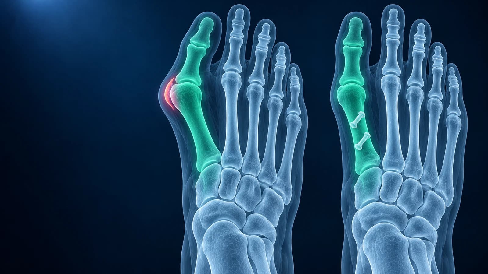
What does a bunion X-ray measure before surgery?
The bump of a bunion is mostly the head of the first metatarsal pushing toward the inner edge of the foot as the bone drifts away from the second metatarsal, while the big toe angles the other way. That is why surgeons look at angles rather than at the bump. The films must be taken standing; a foot photographed lying down can look several degrees straighter.
- Hallux valgus angle (HVA): toe relative to the first metatarsal. A commonly used grading treats up to about 20° as mild, 20° to 40° as moderate and above 40° as severe
- Intermetatarsal angle (IMA): first metatarsal relative to the second. Up to about 11° is mild, 11° to 16° moderate and above 16° severe
- Sesamoid position: whether the two small bones under the big toe joint have slid out toward the second toe
- Distal metatarsal articular angle: the tilt of the joint surface on the metatarsal head, which some surgeons correct separately
- Joint congruency and arthritis: whether the big toe joint surfaces still line up and whether the cartilage space is narrowed
The way each angle is drawn, and why measurements from two readers can differ by a few degrees, is covered in foot X-ray angles explained. If the joint shows narrowing, bone spurs on top and pain on bending the toe upward, the problem may be hallux rigidus rather than, or as well as, a bunion, and that changes the operation.
How does the before X-ray shape the choice of operation?
There are well over a hundred described bunion procedures, but most fall into a few families, and the angles steer the choice in broad terms. A mild deformity with a modest intermetatarsal angle is often treated with a distal osteotomy, such as a chevron cut near the metatarsal head that slides the head outward. Larger angles call for a cut along the shaft, such as a scarf osteotomy, or near the base of the metatarsal, which can swing the bone further.
When the joint where the first metatarsal meets the midfoot is unstable, or the deformity is severe, surgeons may fuse that joint instead, a procedure usually called a Lapidus or first tarsometatarsal fusion. A small wedge cut in the big toe bone itself, an Akin osteotomy, is often added to straighten the toe. If the big toe joint is badly arthritic, fusing that joint is usually more reliable than realigning it. Minimally invasive versions of several of these operations use small incisions and screws placed under X-ray guidance; the post-operative film can look quite different from an open repair while doing the same job.
What should a normal X-ray after bunion surgery look like?
The first post-operative film, often taken in theatre or at the first clinic visit, confirms the correction and the position of the hardware. Expect to see one or two screws, a small plate or staples crossing a sharp, thin cut in the bone, the metatarsal head shifted outward, and the big toe lined up more straight. The two angles are usually re-measured and reported. Soft-tissue swelling around the joint is normal and can make the inner border of the foot still look full.
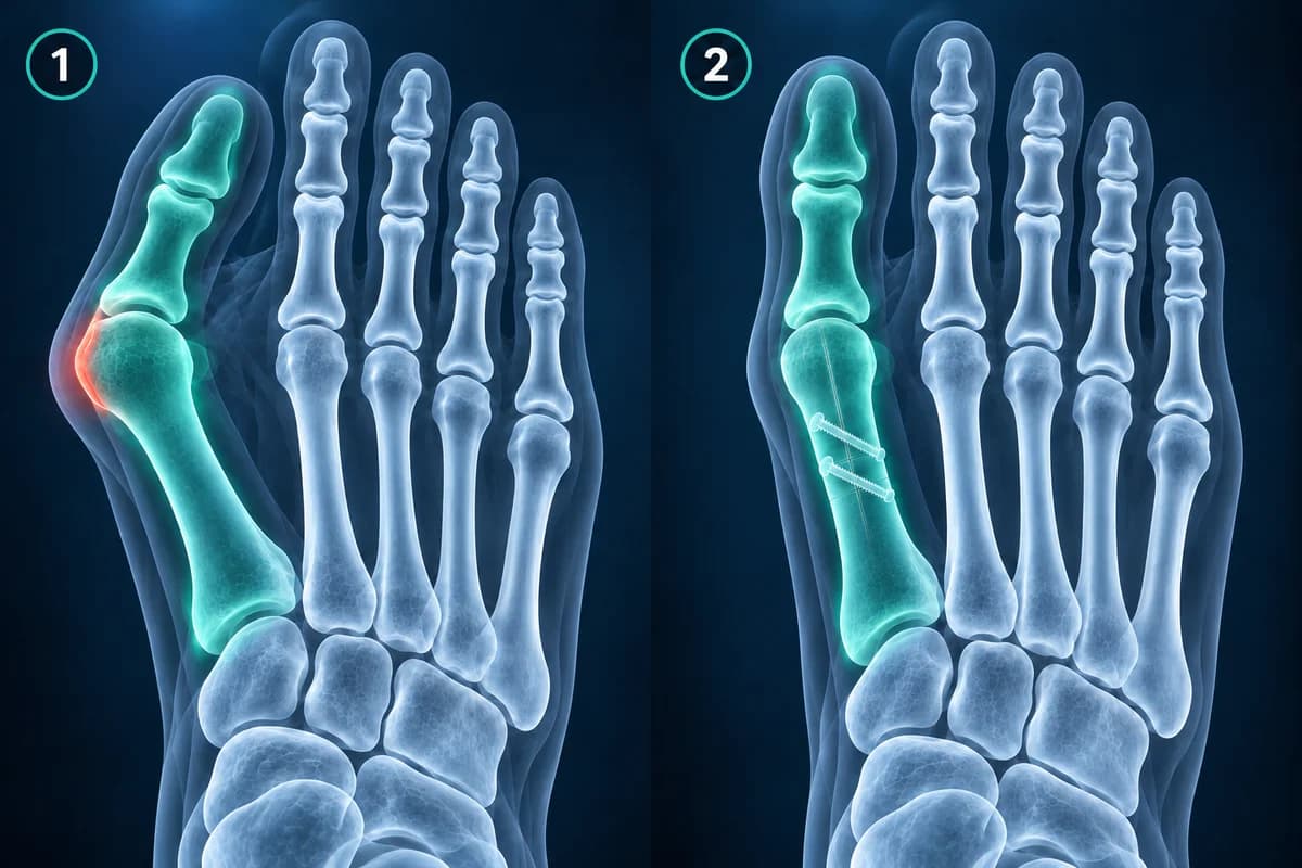
Around six weeks, the cut should look softer and partly bridged by new bone, and many surgeons allow a move from a post-operative shoe into a wide normal shoe if the film and the foot agree. Fusions such as the Lapidus procedure often take longer, sometimes eight to twelve weeks or more, before the joint is judged solid. Later films, if any, mainly confirm that the correction has held. Swelling commonly persists for six months or longer, which is why final results are judged much later than the first X-ray.
If you have the image files, you can compare your pre-operative and post-operative films side by side in the free foot X-ray viewer, which opens them in your browser without uploading. The guide to reading a foot X-ray explains the standard views and landmarks.
What do post-operative bunion X-ray reports mean?
- "Status post distal first metatarsal osteotomy with two-screw fixation" — a description of the operation that was done and the hardware holding it
- "Hardware intact, no periprosthetic lucency" — the screws are unbroken and there is no dark halo around them that would suggest loosening or infection
- "Osteotomy site with interval healing" — new bone is filling the cut compared with the previous film
- "Maintained correction of hallux valgus" — the angles have not drifted back since the last visit
- "Lateral sesamoid subluxation persists" — the sesamoids are still partly off-centre, which some surgeons link with a higher chance of the bunion returning
- "Increased density and flattening of the first metatarsal head" — a pattern that can indicate avascular necrosis, an uncommon complication after distal osteotomies that the surgeon will want to see
Which complications can an X-ray after bunion surgery show?
Most post-operative films are routine. The findings that change management are a loss of correction, where the fragment has shifted or the angles have crept back up; recurrence of the deformity over months to years; overcorrection into hallux varus, where the big toe now points inward; delayed union or nonunion, which is more of a concern after fusions than after osteotomies; broken, backed-out or prominent screws; and changes in the metatarsal head that suggest its blood supply was disturbed. Pain under the ball of the foot after surgery, sometimes called transfer metatarsalgia, may relate to shortening or elevation of the first metatarsal, and can overlap with problems such as a plantar plate tear at the second toe.
Some of these are clear on an X-ray and some are not. Infection, nerve irritation and stiffness are clinical findings first, and early nonunion can need CT to confirm. A worrying phrase on a report is a reason to ask the surgeon what it means for your plan, not a conclusion in itself.
When should you contact your surgeon urgently?
- Redness spreading from the wound, discharge, a wound that opens, or fever
- A toe that turns pale, blue or cold, or loses feeling
- Calf pain, calf swelling or sudden breathlessness in the weeks after surgery
- A sudden increase in pain, a crunch or a visible change in toe position after a slip or knock
- Pain that is getting steadily worse rather than better after the first two weeks
When do bunion images need more than an X-ray?
Before extending a bunion imaging work-up, note that ACR chronic-foot-pain criteria start with X-rays for adults with unexplained persistent pain. Suspected bone or joint disease unresolved by radiographs may warrant MRI or CT without contrast.
Frequently Asked Questions
Why is my foot still wide on the after X-ray?
Early post-operative films include swelling of the soft tissues, which can make the forefoot look wide even when the bones are well corrected. Bunion surgery also aims to realign bones rather than to narrow the foot as such. Compare the bone angles and the position of the metatarsal head rather than the outline of the skin.
Do the screws need to come out after bunion surgery?
Usually not. Screws and plates are commonly left in place for life. They are removed when they irritate the skin or shoe, back out, or break, which is a decision made with the surgeon after the bone has healed.
What angle counts as a successful bunion correction?
There is no single pass mark. Surgeons generally aim to bring the hallux valgus angle under about 15° and the intermetatarsal angle under about 9°, with the sesamoids centred, but a comfortable, stable foot matters more than a number. A few degrees of difference between two readers measuring the same film is normal.
Can a bunion come back after surgery?
Yes, recurrence is a recognised outcome, more often when the original deformity was large, when the sesamoids remain off-centre, or when the first ray is unstable. Follow-up X-rays help detect gradual drift. Whether a recurrence needs treatment depends on symptoms, not only on the angle.
Can AI measure my bunion angles?
An AI explanation can describe the alignment visible on your images and put report terms into plain language, which can help you prepare for a consultation. It is not a diagnosis or a surgical plan; measurements used to decide on surgery are made by the radiologist and surgeon on calibrated standing films.
Sources
Related Articles
What the numbers on your foot X-ray mean: hallux valgus angle, intermetatarsal angle, Meary's angle, calcaneal pitch and Böhler's angle, with normal ranges and how each is measured.
A plain-language guide to your foot X-ray: the three standard views, how to trace alignment from heel to toes, and what fractures, fleck signs, accessory bones and arthritis look like.
How long a broken metatarsal takes to heal week by week, why the fracture line lingers on X-ray, which fractures heal slowly, and what follow-up reports mean.
Related Conditions
Understand your imaging before your next appointment
MRI, X-ray or CT from a CD, folder or ZIP. A PDF report or photos of the films work too.
Free to open and preview, no account needed. An optional AI report from $10 explains your images in plain language, with questions for your doctor.
Your raw files stay on this device. Only rendered images and minimal context are sent when you request an AI report.
Medical Disclaimer: This page is for informational and educational purposes only. It does not constitute medical advice, diagnosis, or treatment. AI-generated analysis may contain errors. Always consult a qualified healthcare professional for medical decisions. Full Disclaimer