
Hallux Rigidus: What Your Big Toe X-ray Says About Stiffness and Arthritis
Hallux rigidus is arthritis of the big toe joint (the first metatarsophalangeal or first MTP joint). On an X-ray it shows up as a dorsal osteophyte, a bone spur on the top of the joint, together with narrowing of the joint space. The spur blocks the toe from bending upward, which is why the joint hurts at push-off. Severity is commonly graded 0 to 4 by combining the X-ray with how far the toe still moves.
Common Symptoms
- Pain at the top of the big toe joint when the toe bends upward during push-off, climbing stairs or squatting
- Gradually shrinking upward movement of the big toe, so it feels jammed or stuck
- A hard bump on the top of the joint that presses against the shoe upper
- Walking on the outer edge of the foot to avoid bending the big toe, with aching on the outside of the foot
- Swelling and stiffness after running, hiking or wearing heels, easing with rest
- Tingling on the top of the big toe where a skin nerve is stretched over the spur
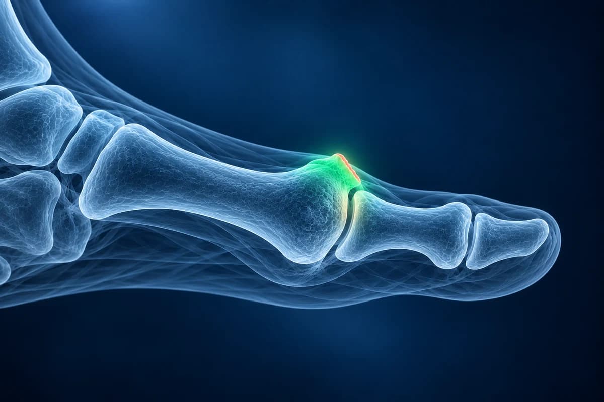
The earlier, milder stage is often called hallux limitus. Below we translate the phrases on a stiff big toe report, explain the grades, and describe when imaging beyond an X-ray helps. This is education, not a diagnosis.
What does “dorsal osteophyte at the first MTP joint” mean?
Dorsal means the top of the foot and an osteophyte is a bony outgrowth at the edge of a joint. In hallux rigidus the spur forms on the top of the first metatarsal head, and sometimes on the base of the toe bone that meets it. It is best seen on the side (lateral) X-ray, where it looks like a beak or lip sticking up from the joint. Each time the toe bends up, the base of the toe jams against the spur, so the joint pinches at the top.
This is a different bump from a bunion. A bunion sits on the inner side of the joint and comes with the big toe drifting toward the second toe; the hallux rigidus spur sits on top and comes with stiffness. The two can exist together, and the bunion and hallux valgus page explains the angles a report uses when they do. The ball-of-foot pain guide compares other symptom locations with a stiff big toe.
What does “joint-space narrowing at the first MTP joint” mean on your X-ray report?
Beyond the spur, a stiff big toe report describes the state of the joint itself. Each phrase below comes with a question worth taking to your clinician.
- “Joint-space narrowing”: the gap between the bones, which on X-ray stands in for the cartilage, is thinner than normal. Ask whether the narrowing is mild or severe, because it is one of the features that sets the grade.
- “Subchondral sclerosis”: the bone just under the cartilage looks whiter and denser because it is taking more load as the cartilage thins. Ask whether it fits the grade suggested for your joint.
- “Subchondral cysts”: small rounded dark spots in that same layer of bone, a feature of more advanced arthritis. Ask whether they affect which operation would be suitable if surgery is ever needed.
- “Flattening of the metatarsal head”: the normally dome-shaped end of the first metatarsal has worn flatter and wider. Ask whether this points to a moderate rather than early grade.
- “Periarticular ossicle” or “loose body”: a small separate piece of bone near the joint, sometimes a broken-off piece of spur, which can catch during movement. Ask whether it explains catching or locking.
- “Osteochondral lesion of the first metatarsal head”: a focal defect in the cartilage and the bone underneath, more often described on MRI and sometimes the starting point of the arthritis in younger people. Ask how large it is and whether it changes the treatment options.
Other wording from foot X-rays is translated in foot X-ray report phrases explained.
How is hallux rigidus graded from 0 to 4?
The grading system most used by foot surgeons was published by Coughlin and Shurnas in 2003. It deliberately blends the X-ray with the examination, because the picture and the symptoms do not always match:
- Grade 0: X-ray normal, mild loss of upward bending, stiffness without real pain.
- Grade 1: a dorsal spur is the main finding with minimal joint narrowing; pain mainly at the end of upward bending.
- Grade 2: spurs, moderate narrowing and some flattening of the metatarsal head; movement more limited and pain more constant at the extremes.
- Grade 3: severe narrowing, larger spurs and possibly cysts; very little upward bending left and near-constant stiffness.
- Grade 4: the same X-ray picture as grade 3, plus pain in the middle of the range when the toe is moved passively, meaning the whole joint surface is involved.
The step between grades 3 and 4 depends on the examination rather than the image, so an X-ray alone can suggest a grade but cannot settle it.
What We Look For on Big Toe Imaging
- Size and position of the dorsal osteophyte on the side view
- How much of the first MTP joint space is preserved, and whether narrowing is uniform or only on top
- Subchondral sclerosis, cysts and flattening of the first metatarsal head
- Loose bodies or ossicles around the joint
- Arthritis or displacement of the sesamoids under the joint
- Coexisting hallux valgus angle that could change surgical choices
Is an X-ray enough, or do you need an MRI for a stiff big toe?
For most people a standing X-ray of the foot, with top-down and side views, is enough to confirm hallux rigidus and grade it. The American College of Radiology Appropriateness Criteria for chronic foot pain start with radiographs, and imaging rarely needs to go further when the X-ray matches the examination. You can review your own films in the free foot X-ray viewer; the side view is where the dorsal spur shows best.
MRI is useful when the X-ray looks normal or near normal but the joint is clearly painful. It can reveal a cartilage defect on the metatarsal head, bone marrow edema (a bruise-like reaction inside the bone), synovitis, or a problem in the sesamoid bones that mimics joint pain. CT is occasionally used to map spurs or cysts before surgery. The trade-offs are compared in foot MRI vs X-ray vs ultrasound.
What causes a big toe joint to stiffen?
Often there is no single cause. Hallux rigidus can follow an injury that damaged the cartilage, such as a forced upward bend in sport (see turf toe), and it can run in families. Repeated flare-ups of gout and inflammatory arthritis can also wear the joint. Foot shape has been debated as a cause, but the evidence is mixed.
How does the grade shape treatment?
Most people start with measures that stop the toe from bending: stiff-soled or rocker-bottom shoes, a carbon insole or a stiff insert under the big toe, avoiding high heels, pain relief and sometimes a joint injection. These do not regrow cartilage but often make walking comfortable.
When pain persists, the grade guides the operation. In the Coughlin and Shurnas series, removing the spur and part of the top of the joint (cheilectomy) worked well for lower grades, while fusion of the joint (arthrodesis) gave reliable pain relief for advanced disease. Other options exist, including osteotomies and joint resurfacing; a foot and ankle surgeon can explain which fits your joint and activities.
When is big toe joint pain urgent?
- The joint becomes suddenly hot, red and exquisitely tender, which suggests gout or infection.
- You have a fever, feel unwell, or there is an open wound near the joint.
- You have diabetes and notice new swelling, warmth or a sore anywhere on the foot.
- The toe looks pale, blue or cold, or you cannot put weight through the foot after an injury.
An AI explanation can help you prepare questions for your clinician. It cannot confirm an injury or its grade on its own and is not a diagnosis.
Frequently Asked Questions
What is the difference between hallux limitus and hallux rigidus?
Both describe a big toe joint that bends upward less than normal. Hallux limitus usually means early, partial loss of motion, while hallux rigidus means advanced arthritis with a large spur and little movement left. Many reports use the terms as two ends of one spectrum.
Can hallux rigidus show up on X-ray before it hurts?
Yes. Spurs and mild narrowing are common incidental findings on foot X-rays taken for other reasons, especially in older adults. A finding without pain or stiffness does not usually need treatment.
Does removing the spur stop the arthritis?
Cheilectomy removes the block to movement and the bump that rubs the shoe, but it does not replace lost cartilage. Arthritis can progress afterward, and some people later need further surgery, which is why it is generally offered for milder grades.
Will I still be able to walk normally after a big toe fusion?
Most people walk normally in suitable shoes after the fusion has healed, which usually takes a few months. The joint no longer bends, so very high heels and activities needing deep toe bending become harder. Your surgeon can set expectations for your sport or work.
Can AI grade hallux rigidus from my X-ray?
Hallux rigidus grading combines X-ray findings such as joint-space loss and bone spurs with pain and movement on examination. AI cannot assign a clinical grade.
Sources
- Coughlin MJ, Shurnas PS. Hallux rigidus: grading and long-term results of operative treatment. J Bone Joint Surg Am. 2003;85(11):2072-2088.
- Expert Panel on Musculoskeletal Imaging. ACR Appropriateness Criteria: Chronic Foot Pain. J Am Coll Radiol.
- Lam A, Chan JJ, Surace MF, Vulcano E. Hallux rigidus: how do I approach it? World J Orthop. 2017;8(5):364-371.
Related Articles
What a bunion X-ray measures before surgery, what a normal post-op X-ray looks like, how long the bone cut takes to heal, and which report findings need attention.
Questions to ask before bunion surgery: what your standing X-ray shows, which operation and why, recovery and weight-bearing, risks, and when not to operate.
What hallux valgus angle, fleck sign, joint space narrowing at the first MTP, pes planus, calcaneal spur and Jones fracture mean on your foot X-ray report.
Understand your imaging before your next appointment
MRI, X-ray or CT from a CD, folder or ZIP. A PDF report or photos of the films work too.
Free to open and preview, no account needed. An optional AI report from $10 explains your images in plain language, with questions for your doctor.
Your raw files stay on this device. Only rendered images and minimal context are sent when you request an AI report.
Medical Disclaimer: This page is for informational and educational purposes only. It does not constitute medical advice, diagnosis, or treatment. AI-generated analysis may contain errors. Always consult a qualified healthcare professional for medical decisions. Full Disclaimer