
Questions to Ask Your Surgeon Before Bunion Surgery
Questions to ask before bunion surgery: what your standing X-ray shows, which operation and why, recovery and weight-bearing, risks, and when not to operate.
Key Takeaways
- Ask to see the measured angles on a standing X-ray, plus sesamoid position and big toe joint arthritis
- Ask which operation family is planned and what would make the surgeon choose another
- Weight-bearing and return to shoes differ a lot between an osteotomy and a Lapidus fusion
- Ask by name about recurrence, transfer pain under the second toe and hallux varus
- A painless bunion that is comfortable in wide shoes usually does not need surgery
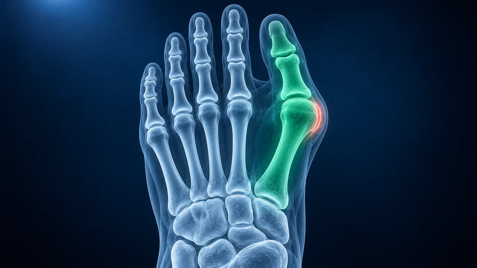
Before bunion surgery, ask four things: what your standing X-ray shows (the hallux valgus angle, normally below 15°, the gap between the first two metatarsals, arthritis and sesamoid position), which operation fits those findings and why, how long until you walk normally in a shoe, and what could go wrong. Surgery is for pain and function, not for the look of the bump.
The questions below are grouped in the order most consultations run. Take the list, your X-ray report and a note of what you most want to be able to do again. This page is a preparation checklist, not advice about whether surgery is right for you.
What does my X-ray show, and was it taken standing?
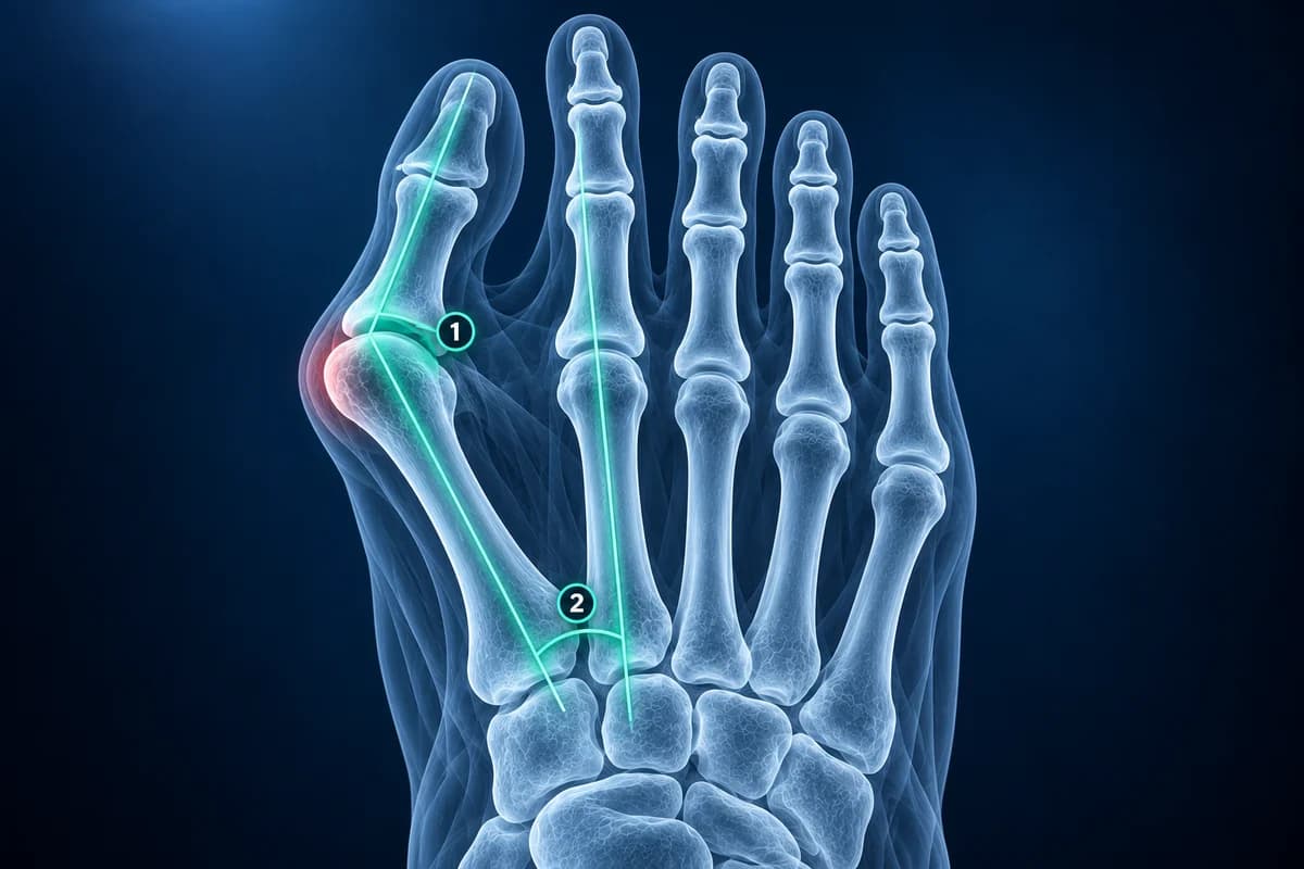
The decision starts with weight-bearing X-rays, because a bunion looks smaller when the foot is not loaded. Ask the surgeon to show you, on the film, the lines they measured.
- What are my hallux valgus and intermetatarsal angles, and is the deformity mild, moderate or severe?
- Have the sesamoids slid out from under the first metatarsal head, and will the operation bring them back?
- Is there arthritis in the big toe joint, and is it the bunion or the arthritis that hurts?
- Is the joint at the base of the first metatarsal loose (hypermobile)?
- Are the second or third toes also involved, for example clawing or a painful joint under them?
How the angles are drawn, and why two readers can differ by a few degrees, is explained in foot X-ray angles explained. If the big toe joint is stiff and narrowed with a spur on top, the main problem may be hallux rigidus, which is treated differently.
Which operation do you recommend for my foot, and why that one?
Bunion operations are named after where the bone is cut or joined. A distal osteotomy cuts near the metatarsal head and suits smaller deformities. A shaft or proximal osteotomy cuts further back and can swing the bone further, for larger angles. A Lapidus procedure fuses the joint at the base of the first metatarsal, often chosen when that joint is loose or the deformity is large. A fusion of the big toe joint itself is usually reserved for severe deformity or arthritis.
- Which of these families is my operation, and what would make you choose another one instead?
- Will you also straighten the toe bone itself or release the tight tissue on the outer side of the joint?
- Do you offer a minimally invasive version, how many have you done, and does it suit my angles?
- Will screws or plates stay in, and how often do they need removing later?
- Would you operate on one foot at a time if both have bunions?
Minimal-access techniques use small incisions and screws placed under X-ray guidance. NICE in the UK found that they work about as well as open surgery, with similar pain and recovery, but advises that only surgeons with specific training and experience in them should do them, so asking about the surgeon’s experience is reasonable rather than rude.
When can I walk, drive and wear normal shoes again?
Recovery depends heavily on the operation. After many distal osteotomies you may walk the same day in a stiff post-operative shoe; after a Lapidus fusion you may be asked to keep weight off the foot for some weeks so the bones can join. Swelling often lasts several months, so a normal shoe may take longer than the wound.
- Can I put weight on the foot straight away, and in what kind of shoe or boot?
- How long before I can drive, given which foot is being operated on?
- How much time off work should I plan for a job on my feet compared with a desk job?
- Will I need blood-clot prevention, physiotherapy or toe exercises?
- When will the follow-up X-rays be, and what will you check on them?
What the after-surgery films should look like, and the phrases their reports use, are covered in bunion surgery X-ray before and after.
What are the risks, and how often do they happen in your hands?
The general risks of any foot operation include infection, wound problems, nerve irritation with numbness near the scar, stiffness and blood clots. Three risks are specific to bunions and are worth asking about by name.
- Recurrence: the toe drifting back over time. Ask what makes it more likely in my foot, such as sesamoids left out of place or a loose first metatarsal joint.
- Transfer pain: load shifting to the ball of the foot under the second toe if the first metatarsal ends up shorter or raised. Ask how the operation protects against that kind of metatarsalgia.
- Hallux varus: over-correction, with the big toe leaning inward. It is uncommon, but ask how it would be managed.
- Non-union: the cut or fused bones not joining. Ask whether smoking or diabetes changes my risk.
Is there a reason not to operate on my bunion now?
Orthopaedic guidance is consistent that a bunion which does not hurt, or which is comfortable in wide shoes, does not need surgery just because the angle is large. Wider toe-box shoes, padding and activity changes are usually tried first. Ask directly whether waiting carries any risk for you, and which symptoms would change the advice. Poor circulation, uncontrolled diabetes, active infection or a foot with reduced feeling can make surgery less safe, so mention them even if they seem unrelated.
If one of the smaller toes is bending into a claw, ask whether it should be corrected at the same time; see hammer toe.
Which symptoms after surgery should make me call straight away?
Ask for a phone number that works outside office hours, and confirm what counts as urgent: fever, increasing redness or discharge from the wound, a calf that is swollen and painful, sudden breathlessness, or a toe that turns pale, blue or very cold. Breathlessness or chest pain needs emergency care, not a call back.
How can I look at my own bunion X-ray before the appointment?
Open the images in our free X-ray viewer, which runs in your browser and does not upload the files. Pick the top-down standing view, use the angle tool along the big toe and first metatarsal, and note anything you want the surgeon to explain. For the phrases in the written report, see foot X-ray report phrases explained.
Frequently Asked Questions
Is bunion surgery worth it?
For people whose bunion hurts despite wider shoes and padding, results are generally good: a review of more than 200 published studies found about 1 in 10 patients dissatisfied afterwards. It is rarely worth it for appearance alone, because recovery takes months and the toe can still drift back.
Is minimally invasive bunion surgery better?
It uses smaller scars and may reduce early swelling, but long-term results look similar to open surgery, it suits some deformities better than others and it depends on the surgeon’s training. Ask whether your angles suit it and what the surgeon’s own results are.
Do I need an MRI before bunion surgery?
Usually not. Standing X-rays show the angles and the joint, which is what the planning needs. MRI or CT is added only for a specific question, such as unexplained pain under another toe or concern about the cartilage.
Can a bunion come back after surgery?
Yes. Recurrence is one of the known risks, more likely when the deformity was severe, the sesamoids stay out of place or the first metatarsal joint was loose. Ask how the chosen operation addresses those factors in your foot.
Can AI help me prepare for a bunion surgery consultation?
AI can explain the angles and phrases in your X-ray report and help you write questions. It cannot examine your foot or tell you which operation to have; that is a decision for you and your surgeon.
Sources
- Ray JJ, Friedmann AJ, Hanselman AE, et al. Hallux Valgus. Foot Ankle Orthop. 2019;4(2):2473011419838500.
- Robinson AH, Limbers JP. Modern concepts in the treatment of hallux valgus. J Bone Joint Surg Br. 2005;87(8):1038-1045.
- Perler A, Dixon M, Alvi F. Hallux Valgus. StatPearls. Treasure Island (FL): StatPearls Publishing.
- American Academy of Orthopaedic Surgeons. Bunions. OrthoInfo.
- National Institute for Health and Care Excellence. Minimally invasive percutaneous surgical techniques with internal fixation for correcting hallux valgus. HealthTech guidance HTG723. 2024.
- Barg A, Harmer JR, Presson AP, et al. Unfavorable Outcomes Following Surgical Treatment of Hallux Valgus Deformity: A Systematic Literature Review. J Bone Joint Surg Am. 2018;100(18):1563-1573.
- Expert Panel on Musculoskeletal Imaging; Said N, Fox MG, Nacey NC, et al. ACR Appropriateness Criteria Chronic Foot Pain: Update 2025. J Am Coll Radiol. 2026;23(4):681-699.
Related Articles
What a bunion X-ray measures before surgery, what a normal post-op X-ray looks like, how long the bone cut takes to heal, and which report findings need attention.
What the numbers on your foot X-ray mean: hallux valgus angle, intermetatarsal angle, Meary's angle, calcaneal pitch and Böhler's angle, with normal ranges and how each is measured.
What hallux valgus angle, fleck sign, joint space narrowing at the first MTP, pes planus, calcaneal spur and Jones fracture mean on your foot X-ray report.
Related Conditions
Understand your imaging before your next appointment
MRI, X-ray or CT from a CD, folder or ZIP. A PDF report or photos of the films work too.
Free to open and preview, no account needed. An optional AI report from $10 explains your images in plain language, with questions for your doctor.
Your raw files stay on this device. Only rendered images and minimal context are sent when you request an AI report.
Medical Disclaimer: This page is for informational and educational purposes only. It does not constitute medical advice, diagnosis, or treatment. AI-generated analysis may contain errors. Always consult a qualified healthcare professional for medical decisions. Full Disclaimer