
Morton's Neuroma: Ultrasound and MRI Findings Explained
Morton's neuroma is a thickened, scarred digital nerve between two metatarsal heads, most often in the third web space. Ultrasound and MRI can both show it as a small oval mass, typically a few millimetres to about a centimetre across. Ultrasound is quick and lets the examiner press on the spot; MRI is better at finding other causes of forefoot pain. Neither replaces the clinical examination.
Common Symptoms
- Burning or sharp pain in the ball of the foot, most often between the third and fourth toes
- A feeling of standing on a pebble or a folded sock that is not there
- Tingling, electric shocks or numbness shooting into the two neighbouring toes
- Pain that worsens in tight, narrow or high-heeled shoes and eases when the shoe comes off
- Relief from rubbing or squeezing the forefoot, followed by return of pain on walking
- A painful click felt between the metatarsal heads when the forefoot is squeezed side to side
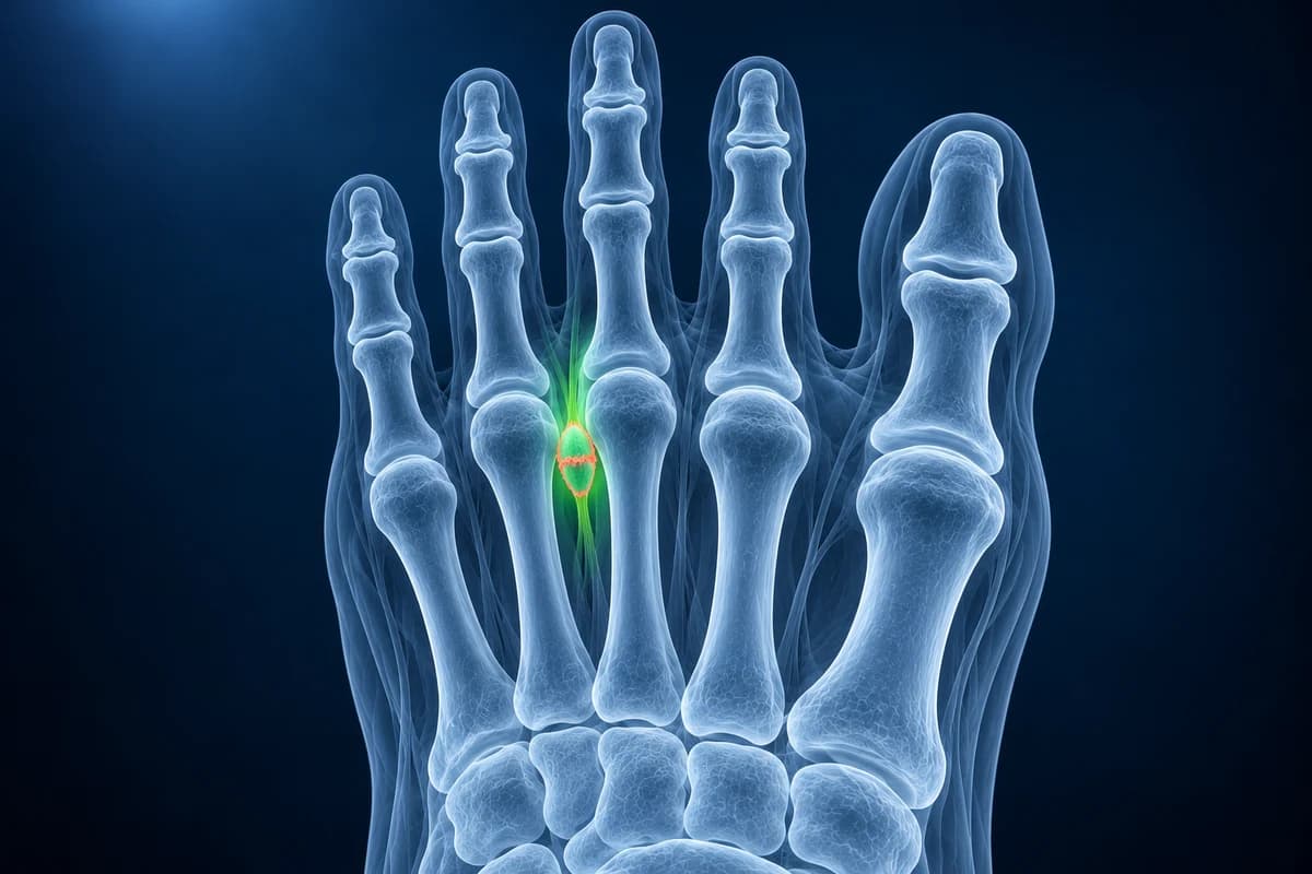
Despite the name, a neuroma here is not a tumour. It is fibrous tissue around a nerve that is repeatedly squeezed between the metatarsal heads and the ligament that ties them together. The sections below explain the words on a neuroma report, how the two scans compare, and why a neuroma seen on a scan is not always the source of pain.
What does “third intermetatarsal space neuroma” on my report mean?
The intermetatarsal spaces, also called web spaces, are numbered from the big toe outward. The third space lies between the third and fourth metatarsals and is where Morton's neuroma is most often found, with the second space next. Reports usually give a size, either the width across the foot or all three dimensions. Two neuromas in one foot, or neuromas in both feet, are not unusual. The foot MRI report glossary helps explain terminology surrounding a web-space finding.
Other phrases that commonly appear:
- “Hypoechoic ovoid mass”: ultrasound wording for a darker-than-surrounding oval lump, the usual look of a neuroma, sometimes with the nerve seen entering one end.
- “Sonographic Mulder sign”: when the examiner squeezes the forefoot, the mass pops downward between the bones, often with a click and a flash of the familiar pain. It supports the diagnosis.
- “Low T1 and intermediate-to-low T2 signal”: MRI wording for the fibrous neuroma, which stays fairly dark on fluid-sensitive images, unlike a fluid-filled bursa or cyst.
- “Intermetatarsal bursitis”: a fluid-filled sac above the neuroma. It can add to pain and often sits alongside a neuroma as a combined bursa and neuroma complex.
- “Enhancing lesion after contrast”: the neuroma took up the contrast dye. Contrast is not needed in every study but can help separate a neuroma from a bursa.
Is ultrasound or MRI better for Morton's neuroma?
Both perform well in experienced hands, and the American College of Radiology Appropriateness Criteria for chronic foot pain list ultrasound and MRI as suitable tests when a neuroma is suspected after the initial X-ray. The choice usually comes down to what else needs ruling out and local expertise.
- Ultrasound is quicker and cheaper, lets the examiner reproduce the pain with pressure, shows the Mulder click in real time and can guide an injection in the same visit. It depends heavily on the skill of the operator.
- MRI shows every web space and every bone at once, so it can catch a plantar plate tear, a metatarsal stress fracture, Freiberg disease or joint inflammation that can feel exactly like a neuroma. Very small neuromas can be hard to see because the foot is scanned lying down.
A longer side-by-side comparison, including cost and what to ask before booking, is in Morton's neuroma ultrasound vs MRI. If you already have an MRI on disc, the free foot MRI viewer opens it in your browser; neuromas are easiest to spot on short-axis slices across the metatarsal heads.
What We Look For in a Neuroma Study
- An oval mass between the metatarsal heads, and which web space it occupies
- Its width across the foot and its relationship to the plantar side of the deep transverse ligament
- Fluid in the intermetatarsal bursa above it and how large that fluid collection is
- Plantar plate integrity at the neighbouring toes, a common mimic in the second space
- Bone marrow edema or a fracture line in the metatarsals that would point to a stress injury
- Joint fluid or synovitis at the lesser toe joints suggesting inflammatory arthritis
Can a neuroma show on a scan without causing pain?
Yes, and this is the most important caveat on any neuroma report. In MRI studies of people with no foot symptoms, Zanetti and colleagues and later Bencardino and colleagues found neuroma-like masses in roughly a third of feet. Zanetti's group also found that small amounts of fluid in the intermetatarsal bursae, up to about 3 mm across, are common and normal in the first three web spaces.
That is why the scan has to be matched to your symptoms. Pain in the same web space where the mass sits, numbness in the two toes that share that nerve, and a positive squeeze test all make a symptomatic neuroma much more likely. A mass in a web space that does not hurt is usually left alone. Some studies suggest that larger neuromas, roughly 5 mm or more across, are more often the ones causing trouble, but size alone does not decide.
Does a plain X-ray help with Morton's neuroma?
An X-ray cannot show the nerve, but it is still the usual first study because it rules out problems that mimic a neuroma: a stress fracture, arthritis of a lesser toe joint, a collapsed second metatarsal head or a toe that is drifting sideways. The guide to reading a foot X-ray shows where to look for each.
What happens after a neuroma is confirmed?
First-line care aims to take pressure off the nerve: wider shoes with a low heel, a metatarsal pad placed just behind the painful spot to spread the bones, and changes to activities that load the forefoot. Corticosteroid injections, often ultrasound-guided, are commonly offered when footwear changes are not enough. Some clinicians also offer other injection or ablation techniques.
Surgery, either releasing the tight ligament above the nerve or removing the affected segment of nerve, is considered when symptoms persist. Removing the nerve leaves permanent numbness between the two toes, and a painful stump neuroma can occasionally form, so it is usually a later option. The size and exact web space on your scan help the surgeon plan the approach.
When should forefoot pain be checked promptly?
- Numbness spreads beyond two neighbouring toes, affects the whole foot, or comes with back pain or leg weakness.
- You have diabetes and new numbness, a wound, warmth or swelling in the foot.
- The forefoot is red, hot and swollen, or you have a fever.
- Pain started after a sudden increase in running or walking and is now sharp on one bone, suggesting a stress fracture.
AI can help explain report wording about a possible neuroma, but it cannot press on your foot, test sensation or decide whether the neuroma is the cause of your pain. Use it to prepare for your appointment. Consider the ball-of-foot pain guide when preparing questions about a possible neuroma.
Frequently Asked Questions
What size of Morton's neuroma is considered significant?
There is no single cut-off. Many symptomatic neuromas measure around 5 mm or more across, but smaller ones can hurt and larger ones can be silent. The match between the location on the scan and where you feel pain matters more than the number.
My ultrasound was normal but my foot still hurts. What next?
A normal ultrasound makes a sizeable neuroma less likely but does not exclude a very small one, and it may miss bone causes. Your clinician may suggest MRI to look for a stress fracture, plantar plate tear or joint inflammation.
Is Morton's neuroma a cancer?
No. It is benign thickening and scarring around a nerve, not a tumour. A mass with an unusual appearance, rapid growth or a location away from the web spaces should be assessed further, as with any lump.
Why does my report mention bursitis as well as a neuroma?
A small fluid sac called the intermetatarsal bursa sits just above the nerve. It often fills with fluid alongside a neuroma, and the two together can form one larger mass on the scan. Small amounts of fluid are normal.
Can AI find a Morton's neuroma on my MRI?
Ultrasound or MRI may show thickening around an interdigital nerve, but similar findings can occur without symptoms. AI cannot confirm that a neuroma is the pain source.
Sources
- Zanetti M, Strehle JK, Zollinger H, Hodler J. Morton neuroma and fluid in the intermetatarsal bursae on MR images of 70 asymptomatic volunteers. Radiology. 1997;203(2):516-520.
- Bencardino J, Rosenberg ZS, Beltran J, Liu X, Marty-Delfaut E. Morton's neuroma: is it always symptomatic? AJR Am J Roentgenol. 2000;175(3):649-653.
- Expert Panel on Musculoskeletal Imaging. ACR Appropriateness Criteria: Chronic Foot Pain. J Am Coll Radiol.
- American Academy of Orthopaedic Surgeons. Morton's Neuroma. OrthoInfo.
Related Articles
What a foot MRI typically costs, why forefoot, midfoot and contrast scans are billed differently, how insurance applies, and how to lower your bill.
Which scan finds a Morton's neuroma better, what each one shows and misses, how reports describe it, and when MRI is worth the extra cost.
What bone marrow edema, plantar fascia thickening, Lisfranc ligament sprain, plantar plate attenuation and sesamoid edema mean on your foot MRI report.
Understand your imaging before your next appointment
MRI, X-ray or CT from a CD, folder or ZIP. A PDF report or photos of the films work too.
Free to open and preview, no account needed. An optional AI report from $10 explains your images in plain language, with questions for your doctor.
Your raw files stay on this device. Only rendered images and minimal context are sent when you request an AI report.
Medical Disclaimer: This page is for informational and educational purposes only. It does not constitute medical advice, diagnosis, or treatment. AI-generated analysis may contain errors. Always consult a qualified healthcare professional for medical decisions. Full Disclaimer