
Gout in the Foot: Big Toe Flares, Tophi and What Imaging Shows
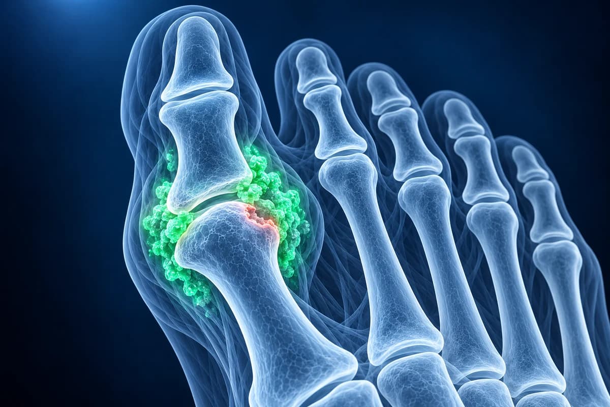
Gout is an inflammatory arthritis caused by urate crystals forming in and around joints when uric acid has been high for a long time. The big toe joint (the first metatarsophalangeal joint) is the classic site, and a flare there is called podagra.
A flare builds over hours, often overnight, to severe pain, heat and swelling, and settles over days to about two weeks. The diagnosis is confirmed by finding crystals in joint fluid; imaging helps when fluid cannot be obtained.
When is a hot, swollen toe not just gout?
A red, hot, swollen joint can also be a joint infection, which can destroy cartilage within days. Get same-day medical care rather than treating it as gout at home if any of the following apply.
- You have a fever, chills or feel generally unwell along with the hot joint
- You have diabetes or numb feet: a hot, swollen foot could be Charcot foot or an infection and needs examination today
- There is a cut, ulcer, draining lump or broken skin over or near the swollen joint
- It is your first attack, or it does not feel like your usual flares
- You have a weakened immune system, a joint replacement, or redness spreading up the foot or leg
This page explains gout in the foot and the words used on X-ray, ultrasound and dual-energy CT reports. It is educational, not a diagnosis, and it does not replace an examination by a clinician.
Common Symptoms
- Sudden, intense pain in the big toe joint, often waking you in the night or early morning
- Shiny red or purplish skin, heat and swelling over the joint
- Pain so sharp that even a bedsheet resting on the toe is hard to bear
- Flares in the instep, midfoot or ankle as well as the big toe
- Firm, painless lumps (tophi) near the toe joints, heel or Achilles area in long-standing gout
- Pain-free spells between flares that get shorter over time if uric acid is not lowered
Why does gout hit the big toe first?
Urate is less soluble at lower temperatures, and the feet are among the coolest parts of the body. The big toe joint also takes heavy load with every step, and joints already affected by wear, such as a stiff toe from hallux rigidus, may be more prone to crystal deposits. Raised uric acid can come from genetics, kidney function, some medicines such as certain diuretics, alcohol (especially beer and spirits), sugary drinks, and diets rich in red meat and seafood. Many people with high uric acid never get gout, so the blood level alone does not make the diagnosis.
A painful big toe has other causes as well. A swollen bursa over a bunion can be red and tender, pain under the joint may come from sesamoiditis, and pseudogout (calcium pyrophosphate crystal arthritis) can mimic a gout flare. In a person with diabetes, a hot foot must always raise the question of Charcot foot or bone infection. The ball-of-foot pain guide places a painful big toe among other forefoot presentations.
How is gout in the foot diagnosed?
The reference test is joint aspiration: a small sample of fluid is drawn from the joint and examined under a polarising microscope, where gout shows needle-shaped crystals. The same sample can be sent for culture to rule out infection. Blood uric acid is useful but can be normal during a flare, so it is often repeated a few weeks later.
When fluid cannot be obtained, the 2015 ACR/EULAR classification criteria accept imaging evidence of urate: the double contour sign on ultrasound or urate deposits on dual-energy CT, along with X-ray erosions typical of gout. X-ray is usually the first image taken, mostly to look for erosions, other arthritis and fractures. To compare the tests, see foot MRI vs X-ray vs ultrasound and how to read a foot X-ray.
What does “punched-out erosion” or “double contour sign” mean on a gout report?
- “Punched-out erosion with overhanging edge”: a rounded bite out of the bone near the joint, with a thin lip of bone curving over it. It is the classic X-ray sign of long-standing gout, usually at the inner side of the first metatarsal head; ask whether it means urate-lowering treatment is now recommended.
- “Joint space relatively preserved”: unlike wear-and-tear arthritis, gout tends to keep the joint gap until late. Ask whether the X-ray shows any osteoarthritis as well as gout.
- “Soft tissue density consistent with tophus”: a lump of packed urate crystals in the soft tissue, sometimes faintly dense on X-ray. Ask what uric acid target would make it shrink.
- “Double contour sign”: on ultrasound, a bright line of urate coating the joint cartilage, parallel to the bone beneath. Ask whether it is enough to confirm gout or whether a joint fluid test is still needed.
- “Urate deposition on DECT”: dual-energy CT colour-codes urate, often green, around tendons and joints, though small artefacts can be coded too. Ask whether the deposits are in a pattern typical of gout.
- “Synovitis” or “effusion with marrow edema” on MRI: active joint inflammation, which can also look like infection. Ask whether infection has been ruled out, ideally with a joint fluid sample.
Early in gout the X-ray is often normal apart from soft tissue swelling, because erosions take years of uncontrolled crystal deposits to form. You can open your images in the free foot X-ray viewer, or load a dual-energy study in the free foot CT viewer. More report wording is explained in foot X-ray report phrases explained.
What We Look For on Imaging
- Juxta-articular erosions with sclerotic margins and overhanging edges at the first metatarsal head
- Asymmetric soft tissue swelling or tophaceous densities around the toe joints, midfoot or heel
- Relatively preserved joint space and bone density compared with the degree of erosion
- Double contour sign, tophi or hyperechoic aggregates on ultrasound reports
- Colour-coded urate deposits around joints and tendons on dual-energy CT
- Features that point elsewhere, such as osteoarthritic spurs, chondrocalcinosis or bone destruction under an ulcer
How is gout in the foot treated?
A flare is treated as early as possible with colchicine, an anti-inflammatory such as naproxen, or a steroid given by mouth or by injection; the choice depends on kidney function, stomach and heart risks, diabetes and other medicines. Rest, elevation and ice help. Which option is safe for you is a decision for your doctor or pharmacist.
The long-term goal is to dissolve the crystals by lowering uric acid. The 2020 ACR guideline strongly recommends urate-lowering therapy, with allopurinol as the usual first choice, for people with one or more tophi, X-ray damage from gout, or two or more flares a year. It recommends a treat-to-target approach with a serum urate below 6 mg/dL (about 360 µmol/L), and flare-preventing medicine for the first 3 to 6 months, because flares can briefly increase when urate starts falling. Erosions do not usually heal, but stable urate stops new crystals forming and tophi shrink over time.
What should I ask my doctor?
- Has infection been ruled out, and would aspirating the joint help confirm gout?
- Does my X-ray show erosions or tophi, and how far has the joint been damaged?
- Should I start urate-lowering treatment, and what is my target uric acid level?
- Could any of my current medicines be raising my uric acid?
- Would ultrasound or dual-energy CT change the plan in my case?
Frequently Asked Questions
Can gout show on a foot X-ray?
Only in long-standing or poorly controlled gout. Early flares usually show nothing more than soft tissue swelling. After years of crystal deposits, X-ray can show punched-out erosions with overhanging edges and tophi, typically at the big toe joint.
What is the double contour sign on ultrasound?
It is a bright line of urate crystals lying on the surface of the joint cartilage, running parallel to the bone surface. It is one of the imaging features counted in the ACR/EULAR gout classification criteria.
Can I have gout with a normal uric acid level?
Yes. Uric acid can fall during a flare, so a normal result at that moment does not rule gout out. Doctors often recheck the level a few weeks after the flare settles, and joint fluid remains the most reliable test.
How can I tell gout from an infected toe?
You often cannot tell from the outside. Fever, a wound near the joint, diabetes, or a first-ever attack are reasons to be examined the same day, and a joint sample checked for crystals and bacteria is what separates the two.
Can AI analysis confirm gout from my scan?
Joint fluid crystals can confirm gout, while examination and testing help distinguish infection. AI cannot confirm gout or rule out an infected joint.
Sources
- FitzGerald JD, Dalbeth N, Mikuls T, et al. 2020 American College of Rheumatology Guideline for the Management of Gout. Arthritis Care Res (Hoboken). 2020;72(6):744-760.
- Neogi T, Jansen TL, Dalbeth N, et al. 2015 Gout classification criteria: an American College of Rheumatology/European League Against Rheumatism collaborative initiative. Arthritis Rheumatol. 2015;67(10):2557-2568.
- Menon SG, Rednam M, Gujarathi R, Maher L. Gout. StatPearls. Treasure Island (FL): StatPearls Publishing.
Related Articles
Why a diabetic foot X-ray comes first, when MRI is used to tell bone infection from Charcot foot, and why scans alone do not decide treatment.
A plain-language guide to your foot X-ray: the three standard views, how to trace alignment from heel to toes, and what fractures, fleck signs, accessory bones and arthritis look like.
Understand your imaging before your next appointment
MRI, X-ray or CT from a CD, folder or ZIP. A PDF report or photos of the films work too.
Free to open and preview, no account needed. An optional AI report from $10 explains your images in plain language, with questions for your doctor.
Your raw files stay on this device. Only rendered images and minimal context are sent when you request an AI report.
Medical Disclaimer: This page is for informational and educational purposes only. It does not constitute medical advice, diagnosis, or treatment. AI-generated analysis may contain errors. Always consult a qualified healthcare professional for medical decisions. Full Disclaimer