
Diabetic Foot Imaging: X-Ray, MRI and What to Expect
Why a diabetic foot X-ray comes first, when MRI is used to tell bone infection from Charcot foot, and why scans alone do not decide treatment.
Key Takeaways
- A hot swollen foot, a new ulcer or fever with diabetes needs same-day care, not a scan first
- Plain X-rays are the first test, even when there was no injury
- Early bone infection and early Charcot can both look normal on X-ray
- MRI is the usual next test and separates the two mainly by location and pattern
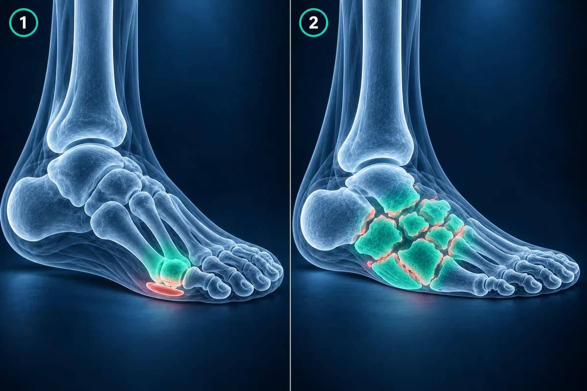
- Infection usually sits under an ulcer; Charcot usually involves several midfoot joints
- Probe-to-bone, blood tests and sometimes a bone biopsy complete the picture
Which diabetic foot problems need care today, before any scan?
If you have diabetes, do not wait for an imaging appointment or an online explanation when your foot shows any of the signs below. Contact your diabetes foot team, GP or urgent care the same day, and keep weight off the foot until you are seen.
- Go to the emergency department now if a foot wound or swollen foot comes with fever, chills, vomiting, feeling confused or generally unwell, very high blood sugars, or redness spreading up the leg.
- Seek same-day care for one foot that is suddenly hotter, redder or more swollen than the other, even if it barely hurts.
- Seek same-day care for any new ulcer, blister or wound, or one that is draining, smells bad, has turned black, or is getting deeper.
- Seek same-day care if the shape of the foot seems to be changing or the arch is dropping, even without pain.

For a diabetic foot problem, a plain X-ray almost always comes first, even when there was no injury. If it is normal or unclear and infection or Charcot foot is still suspected, MRI is usually the next test. The American College of Radiology notes that about 20% of people with a diabetic foot ulcer go on to develop bone infection, which is why scans are ordered early.
This guide explains what to expect from each test, the report phrases you are likely to read, and why the scans are only one part of the decision. It is educational and not a diagnosis. Diabetic foot problems are managed by a clinical team who examine the foot in person.
Why is an X-ray the first test for a diabetic foot?
The 2025 ACR Appropriateness Criteria for suspected osteomyelitis of the foot in diabetes recommend X-rays of the foot as the first imaging test, and the IWGDF/IDSA infection guideline also starts with plain X-rays. They are quick, cheap and widely available, and they give a baseline: bone loss, a fracture, joint destruction, gas in the soft tissues, a foreign body or arthritis that changes what the next step should be.
When nerve damage has reduced feeling in the foot, there may be no memorable injury at all. A warm, swollen foot with a normal shape still deserves an X-ray, ideally standing if that is safe, because the IWGDF Charcot guideline uses X-rays of the foot and ankle as the first step in suspected active Charcot foot. A clear X-ray is a useful starting point to compare against later films.
What can a diabetic foot X-ray miss?
Early bone infection and early Charcot foot can both look normal on X-ray. According to the ACR, infection usually has to destroy 30% to 50% of the bone's mineral before X-ray shows it, so the first films may look normal for 10 to 14 days or longer while the infection is already active. The IWGDF Charcot guideline likewise notes that active Charcot foot can show on MRI while the X-rays still look normal. That is why a normal X-ray does not end the workup when the clinical picture still points to infection or Charcot, and why clinicians often repeat X-rays over the following weeks.
Phrases you may see on an X-ray report include "cortical erosion" or "periosteal reaction" near a toe or metatarsal head, which raise concern for bone infection; "soft tissue gas", which is urgent; and "fragmentation", "subluxation" or "collapse of the midfoot", which point toward Charcot. Ask the clinician which of these apply to you and whether further imaging will change the plan. You can look through your own films in the free X-ray viewer.
When is MRI added, and how does it tell infection from Charcot?
According to the ACR, when the X-rays are negative or unclear, or when they already show infection and surgery needs planning, MRI of the foot is the most common next test. MRI shows bone marrow edema, fluid collections, sinus tracts and soft-tissue infection long before X-ray changes appear. A contrast injection is often given to show abscesses and dead tissue more clearly.
Both bone infection and Charcot foot cause marrow edema, so radiologists read the pattern rather than the signal alone. A review in RadioGraphics describes the key clues: infection usually starts from the skin, so it sits in bone directly beneath an ulcer, often at the toes, the metatarsal heads or the heel, with a track or abscess in between. Charcot foot usually affects several joints in the midfoot, centered on the joints themselves, often without a nearby ulcer. Read more about each on the diabetic foot osteomyelitis and Charcot foot pages.
What does "marrow edema adjacent to the ulcer" mean on an MRI report?
It means the bone under the wound holds extra fluid, which can be infection inside the bone or a reaction to the nearby soft-tissue infection. Radiologists then look at the T1 images: a confluent area of "low T1 marrow signal", where the normal fatty marrow is fully replaced, is the pattern most strongly linked to osteomyelitis, while a hazy, net-like pattern is more often reactive. Edema with normal T1 signal is not automatically harmless: the ACR notes that when it sits next to an ulcer, abscess or sinus tract, bone infection is still likely. Ask whether the report says "consistent with" or only "cannot exclude" osteomyelitis, because the two lead to different conversations.
For Charcot, typical phrases are "subchondral marrow edema at the tarsometatarsal joints", "neuropathic arthropathy" and "joint effusions with fragmentation". A "sinus tract" or "abscess" on a Charcot foot means infection is present as well and needs urgent attention. Our guide to reading a foot MRI explains the sequences these phrases refer to.
Why does imaging alone not decide treatment?
Scans describe what the bone looks like, not what is growing in it. The IWGDF/IDSA guideline recommends combining a probe-to-bone test at the bedside, plain X-rays and a blood inflammatory marker, then adding MRI when the diagnosis is still in doubt. When the answer remains unclear or the choice of antibiotic matters, a bone sample taken for culture is the most reliable way to confirm infection and identify the germ.
Infection and Charcot can also occur in the same foot, and MRI changes after surgery or a recent fracture can look similar to both. Treatment decisions also depend on things a scan cannot show well: blood flow to the foot, how deep the wound probes, blood sugar control and how the foot is being offloaded. That is why the guidelines place these decisions with a multidisciplinary foot team rather than with a single report.
How quickly should a suspected Charcot foot be seen?
Quickly. NICE advises that anyone with suspected Charcot arthropathy be referred to a multidisciplinary foot care service within one working day, with a weight-bearing X-ray of the foot and ankle and an MRI if the X-ray is normal but the suspicion remains. The IWGDF Charcot guideline recommends starting knee-high offloading straight away while the tests are done, rather than waiting for the MRI result. A skin temperature difference of about 2 °C between the feet has often been used as a sign of active disease, but the guideline stresses that a warmer foot alone can neither confirm nor rule out Charcot.
Are other scans ever used?
Sometimes. People who cannot have MRI, for example because of certain implants, or whose foot has metal from previous surgery, may be offered nuclear medicine tests such as a labeled white blood cell scan, or CT to look at bone destruction and gas. Ultrasound can guide drainage of a collection. These are chosen case by case; MRI remains the usual second step after X-ray.
Frequently Asked Questions
Can an X-ray show osteomyelitis in a diabetic foot?
Sometimes, once bone has been destroyed enough to show as erosion or a periosteal reaction. Early infection is often invisible, so a normal X-ray does not rule it out when there is a deep ulcer or the probe reaches bone.
Is MRI or a bone scan better for a diabetic foot infection?
The ACR lists MRI as the usual next test after X-ray. Nuclear medicine scans are kept for people who cannot have MRI or when metal in the foot spoils the images.
Can MRI tell Charcot foot from infection?
Often, using where the changes sit and whether there is an ulcer, sinus tract or abscess nearby. When both are present or the pattern is mixed, the clinical exam, blood markers and sometimes a bone biopsy decide.
Why was I told to stay off my foot before the MRI?
If active Charcot foot is possible, walking on the softened bones can make the foot collapse. Guidelines advise offloading straight away and not waiting for the scan result.
Will I need contrast for a diabetic foot MRI?
Often, because contrast helps show abscesses and dead tissue. It needs reasonable kidney function, which is checked first in people with diabetes. Your imaging team decides whether it is needed.
Sources
- Expert Panel on Musculoskeletal Imaging; Baker JC, Northrup BE, Ahlawat S, et al. ACR Appropriateness Criteria Suspected Osteomyelitis of the Foot in Patients With Diabetes Mellitus: 2025 Update. J Am Coll Radiol. 2025;22(11S):S713-S737.
- Senneville É, Albalawi Z, van Asten SA, et al. IWGDF/IDSA guidelines on the diagnosis and treatment of diabetes-related foot infections (IWGDF/IDSA 2023). Diabetes Metab Res Rev. 2024;40(3):e3687.
- Wukich DK, Schaper NC, Gooday C, et al. Guidelines on the diagnosis and treatment of active Charcot neuro-osteoarthropathy in persons with diabetes mellitus (IWGDF 2023). Diabetes Metab Res Rev. 2024;40(3):e3646.
- Donovan A, Schweitzer ME. Use of MR imaging in diagnosing diabetes-related pedal osteomyelitis. RadioGraphics. 2010;30(3):723-736.
- Johnson PW, Collins MS, Wenger DE. Diagnostic utility of T1-weighted MRI characteristics in evaluation of osteomyelitis of the foot. AJR Am J Roentgenol. 2009;192(1):96-100.
- National Institute for Health and Care Excellence. Diabetic foot problems: prevention and management (NG19).
Related Articles
A plain-language guide to your foot MRI: how T1, fluid-sensitive and STIR images differ, which plane shows what, and what bone marrow edema, plantar plate tear and neuroma mean in your report.
A plain-language guide to your foot X-ray: the three standard views, how to trace alignment from heel to toes, and what fractures, fleck signs, accessory bones and arthritis look like.
X-ray, MRI, ultrasound or CT for foot pain? What each test shows and misses, which one usually comes first, and how the choice changes for stress fractures, neuromas, plantar fasciitis, Lisfranc injuries and the diabetic foot.
Related Conditions
Understand your imaging before your next appointment
MRI, X-ray or CT from a CD, folder or ZIP. A PDF report or photos of the films work too.
Free to open and preview, no account needed. An optional AI report from $10 explains your images in plain language, with questions for your doctor.
Your raw files stay on this device. Only rendered images and minimal context are sent when you request an AI report.
Medical Disclaimer: This page is for informational and educational purposes only. It does not constitute medical advice, diagnosis, or treatment. AI-generated analysis may contain errors. Always consult a qualified healthcare professional for medical decisions. Full Disclaimer