
Foot MRI vs X-ray vs Ultrasound: Which Scan Do You Need?
X-ray, MRI, ultrasound or CT for foot pain? What each test shows and misses, which one usually comes first, and how the choice changes for stress fractures, neuromas, plantar fasciitis, Lisfranc injuries and the diabetic foot.
Key Takeaways
- A standing X-ray is the usual first test for foot pain and the reference for alignment
- Ultrasound suits one focused soft-tissue question and allows scanning while the foot moves
- MRI is the most sensitive test for stress fractures, bone infection and unclear pain
- CT maps complex fractures and bony coalitions in three dimensions
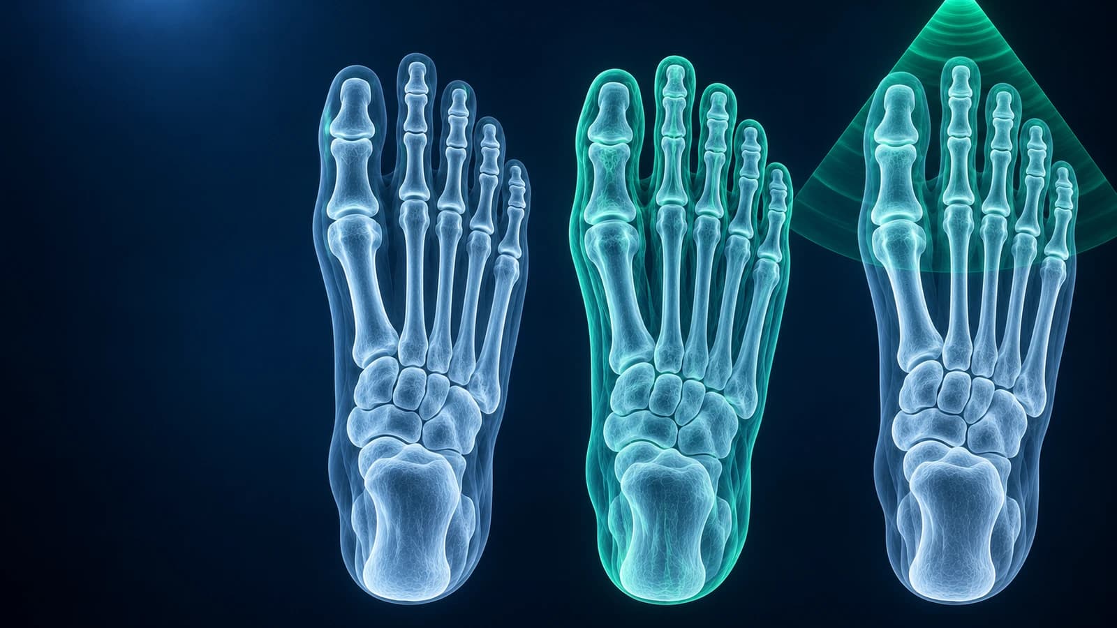
For most foot pain, a standing X-ray comes first because it is quick, cheap and shows fractures, alignment and arthritis. Ultrasound is the usual next step for a single soft-tissue target such as a Morton’s neuroma or the plantar fascia, MRI when the cause is unclear or a stress fracture or infection is suspected, and CT when a complex fracture needs mapping. This order follows the American College of Radiology Appropriateness Criteria.
The best scan is the one that answers the question your clinician is asking. Below, each test is compared on what it sees, what it misses and where it fits, followed by the usual choice for the most common foot problems. This is general education, not a referral; your own pathway depends on your exam and history.
What does a foot X-ray show, and what does it miss?
X-ray is a shadow picture of bone. Taken standing, it is the reference test for bunion angles, arch shape and midfoot alignment, and it reliably shows most acute fractures, dislocations, arthritis, gout erosions, accessory bones and metal from earlier surgery. It uses a small dose of radiation and takes minutes. Its blind spots are soft tissue (fascia, tendons, nerves, the plantar plate) and early bone stress, which may take two to three weeks to appear. Our guide on how to read a foot X-ray explains the views and report terms.
When is ultrasound better than MRI for the foot?
Ultrasound shines when the problem is small, near the skin and has a clear location. The examiner can press on the sore spot, move the toe and compare with the other foot during the scan, which MRI cannot do. Typical uses are a neuroma between the metatarsal heads, a thickened plantar fascia, a plantar plate tear checked while the toe is moved, fluid in a joint or bursa, a ganglion, and a splinter or other foreign body that X-ray cannot see. It also guides injections precisely. It involves no radiation and is usually cheaper than MRI.
The trade-offs: ultrasound cannot see inside bone, so bone marrow edema and most stress injuries are invisible; it shows only the area scanned; and the result depends heavily on the operator’s experience. Pooled studies for Morton’s neuroma found ultrasound and MRI similarly sensitive in experienced hands; for plantar plate tears ultrasound was more sensitive but MRI more accurate overall, so the choice depends on the question, local expertise and cost.
What can a foot MRI show that the others cannot?
MRI sees inside bone and through all soft tissues at once, without radiation. That makes it the most sensitive test for early stress fractures and bone bruises, the preferred test when several structures could be the cause, and the key test when bone infection is suspected in a diabetic foot. It can show cartilage damage, small ligament tears, tendon disease and the full extent of a mass. The downsides are cost, waiting times, lying still for the length of the scan, and a field of view that may cover only the forefoot or only the hindfoot. Its very sensitivity can also report incidental findings that are not the cause of pain. See how to read a foot MRI and what drives foot MRI cost.
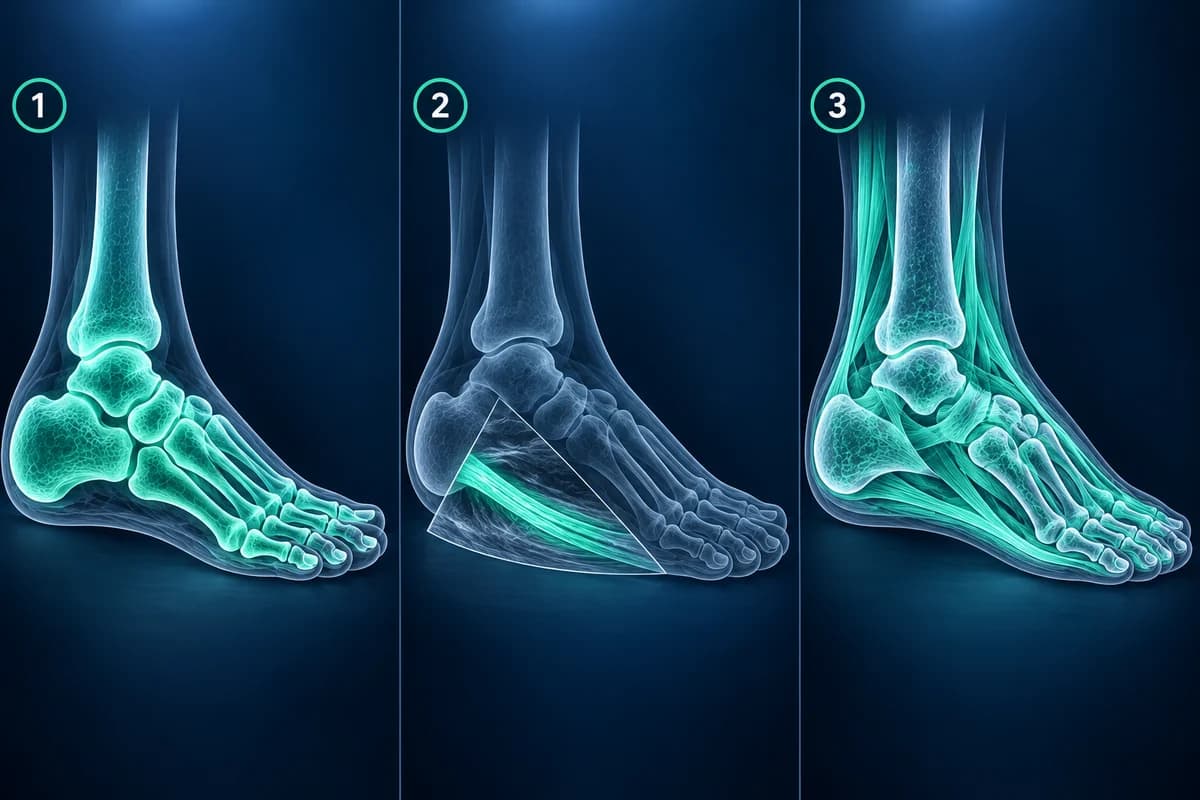
When is a CT scan of the foot used?
CT is a three-dimensional X-ray. It does not replace MRI for soft tissue but beats every other test at showing the exact shape of a fracture. It is the usual follow-up for a heel bone break, a midfoot fracture-dislocation, a fracture into a joint surface, and a bony tarsal coalition before surgery. Some centres offer weight-bearing CT, which shows alignment under body weight in three dimensions. CT uses more radiation than plain X-ray, though foot CT doses are low because the scan avoids the body trunk.
Which scan is used for common foot problems?
- Suspected metatarsal stress fracture: X-ray first; if it is normal and suspicion stays high, MRI or a repeat X-ray after a couple of weeks.
- Burning pain between the toes from a Morton’s neuroma: often diagnosed by examination; ultrasound or MRI if confirmation is needed. Our article on neuroma ultrasound versus MRI goes deeper.
- Heel pain typical of plantar fasciitis: usually no scan at first; X-ray, ultrasound or MRI if pain persists or the story is unusual.
- Midfoot twist with swelling and bruising on the sole: X-ray, ideally standing or comparing both feet, then CT or MRI if a Lisfranc injury is still suspected.
- Big toe bump or stiffness: standing X-ray, which measures bunion angles and shows joint wear; other scans are rarely needed.
- Diabetic foot with an ulcer and possible bone infection: X-ray first, then MRI; nuclear medicine scans are an option when MRI is not possible. See diabetic foot osteomyelitis.
Should I ask for an MRI straight away?
Usually not. Guidelines place MRI after X-ray for most foot complaints because the X-ray answers many questions on its own, changes what the MRI is looking for, and is needed to interpret some MRI findings correctly. An MRI done too early or for the wrong question can show changes that were already there before the pain started. If your symptoms are not improving after a reasonable period of treatment, or an X-ray has not explained them, ask your clinician whether the next test should be MRI, ultrasound or CT and what result would change the plan.
When should foot pain be seen urgently instead of waiting for a scan?
Go to urgent care the same day if you cannot bear weight after an injury, the foot looks deformed, a toe is pale, blue or cold, there is an open wound over a possible fracture, or a foot affected by diabetes becomes hot, red and swollen, with or without fever. In these cases the right test is ordered by the treating team on the spot.
How can I look at my own foot scans?
X-ray, CT and MRI discs usually contain DICOM files you can open yourself. Use the free X-ray viewer for plain films, the free MRI viewer for MRI series and the free CT viewer for CT. All of them open the files in your browser without uploading the originals. Ultrasound is usually saved as a few still images, so the written report carries most of its value.
Frequently Asked Questions
Is MRI or ultrasound better for Morton’s neuroma?
Both detect most neuromas when performed by experienced operators. Ultrasound is cheaper and lets the examiner press on the web space during the scan; MRI covers the whole forefoot and can reveal other causes of pain at the same time.
Can ultrasound detect a foot stress fracture?
Ultrasound can sometimes show swelling over the bone surface at a stress fracture, but it cannot see inside the bone. MRI remains the most sensitive test when X-ray is normal and a stress fracture is still suspected.
Why do I need an X-ray if I am already getting an MRI?
The X-ray shows standing alignment, calcification and bone detail that MRI may not, and radiologists use it to interpret the MRI correctly. Many guidelines recommend having one before MRI for that reason.
Is a foot CT scan a lot of radiation?
Foot CT exposes far less of the body than a chest or abdominal CT, so the dose is low compared with most CT scans, though higher than a plain X-ray. It is used when the extra fracture detail will change treatment.
Can AI help me decide which foot scan I need?
AI can explain what an existing scan shows and which questions might remain, but the decision to order a test belongs to the clinician who examined you. Bring the explanation to that conversation.
Sources
- Expert Panel on Musculoskeletal Imaging; Said N, Fox MG, Nacey NC, et al. ACR Appropriateness Criteria Chronic Foot Pain: Update 2025. J Am Coll Radiol. 2026;23(4):681-699.
- Expert Panel on Musculoskeletal Imaging; Gorbachova T, Chang EY, Ha AS, et al. ACR Appropriateness Criteria Acute Trauma to the Foot. J Am Coll Radiol. 2020;17(5S):S2-S11.
- Bignotti B, Signori A, Sormani MP, et al. Ultrasound versus magnetic resonance imaging for Morton neuroma: systematic review and meta-analysis. Eur Radiol. 2015;25(8):2254-2262.
- Albright RH, Brooks BM, Chingre M, et al. Diagnostic accuracy of magnetic resonance imaging (MRI) versus dynamic ultrasound for plantar plate injuries: a systematic review and meta-analysis. Eur J Radiol. 2022;152:110315.
- Expert Panel on Musculoskeletal Imaging; Baker JC, Northrup BE, Ahlawat S, et al. ACR Appropriateness Criteria Suspected Osteomyelitis of the Foot in Patients With Diabetes Mellitus: 2025 Update. J Am Coll Radiol. 2025;22(11S):S713-S737.
Related Articles
A plain-language guide to your foot MRI: how T1, fluid-sensitive and STIR images differ, which plane shows what, and what bone marrow edema, plantar plate tear and neuroma mean in your report.
A plain-language guide to your foot X-ray: the three standard views, how to trace alignment from heel to toes, and what fractures, fleck signs, accessory bones and arthritis look like.
Which scan finds a Morton's neuroma better, what each one shows and misses, how reports describe it, and when MRI is worth the extra cost.
Related Conditions
Understand your imaging before your next appointment
MRI, X-ray or CT from a CD, folder or ZIP. A PDF report or photos of the films work too.
Free to open and preview, no account needed. An optional AI report from $10 explains your images in plain language, with questions for your doctor.
Your raw files stay on this device. Only rendered images and minimal context are sent when you request an AI report.
Medical Disclaimer: This page is for informational and educational purposes only. It does not constitute medical advice, diagnosis, or treatment. AI-generated analysis may contain errors. Always consult a qualified healthcare professional for medical decisions. Full Disclaimer