
Lisfranc Injury: Fleck Sign, Midfoot Diastasis and What Your Imaging Shows
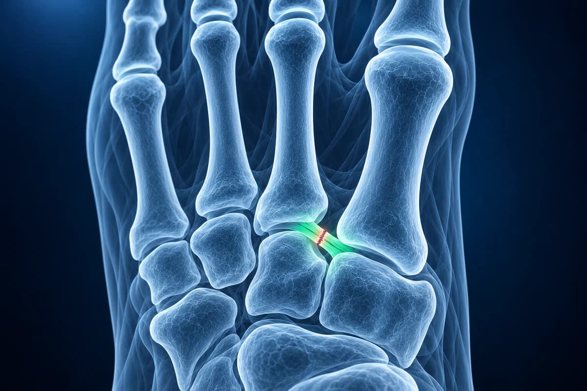
A Lisfranc injury is damage to the ligaments or bones of the tarsometatarsal joints in the middle of the foot. It is easy to miss: a non-weight-bearing X-ray can look normal, so a widening of more than about 2 mm between the first and second metatarsal bases, or a tiny "fleck sign", is often only visible on standing views, CT or MRI.
Some signs described on this page need prompt medical care. If you have them, get care first. Reading about imaging can wait.
Common Symptoms
- Pain across the top of the midfoot that worsens when standing on tiptoe or pushing off
- Bruising on the sole of the arch (plantar ecchymosis), a classic warning sign of a tarsometatarsal injury
- Swelling over the midfoot that seems out of proportion to a "simple sprain"
- Tenderness directly over the joint between the first and second metatarsal bases
- Pain when the forefoot is gently twisted while the heel is held still
- Difficulty bearing weight, or a midfoot that still hurts with walking weeks after the injury
When is midfoot pain an emergency?
Go to an emergency department now if you cannot take four steps on the foot, if the midfoot looks deformed, or if bone or a wound is exposed. High-energy Lisfranc fracture-dislocations from falls, car crashes or crush injuries can cause compartment syndrome: severe, bursting pain that keeps rising despite painkillers, a tense swollen foot, pain when the toes are passively stretched, numbness, or toes that turn pale or cold. These signs need same-day surgical assessment, not a scan review.
What is the Lisfranc joint and why does it matter?
The tarsometatarsal joint complex is the row of joints where the five long metatarsal bones meet the three cuneiforms and the cuboid. The base of the second metatarsal sits recessed between the medial and lateral cuneiforms like a keystone in an arch. There is no strong ligament tying the first and second metatarsal bases together; instead, the Lisfranc ligament anchors the second metatarsal base to the medial cuneiform. When that ligament fails, the keystone loosens and the arch can slowly collapse under body weight.
Injuries range from a subtle sprain in a football player who was landed on while the foot was pointed down, to a violent fracture-dislocation in a motorbike crash. The low-energy, purely ligamentous version is the one that slips through: the foot is swollen, the first X-ray is called normal, and the patient is told it is a sprain. Because the midfoot transmits load with every step, an unstable injury that heals out of place can leave long-term pain and post-traumatic midfoot arthritis, which is why getting the stability question answered early matters.
Why was my Lisfranc injury missed on the first X-ray?
Emergency foot X-rays are usually taken with the patient sitting or lying, because standing hurts. Without body weight pushing the bones apart, a torn but undisplaced Lisfranc ligament lets the joint sit in a near-normal position. The gap that gives the injury away often opens only when the foot is loaded. Overlapping bones on the lateral view and swelling that hides fine detail add to the problem.
That is why the ACR Appropriateness Criteria for acute foot trauma recommend weight-bearing views when a midfoot or Lisfranc injury is suspected, with a comparison view of the other foot, and CT or MRI when standing films are normal but suspicion stays high. If you were told your foot was "just sprained" and the arch still hurts to stand on after a week or two, ask whether standing films have been done. Our guide to Lisfranc injuries missed on X-ray walks through the views in more detail.
What do Lisfranc report phrases mean?
- "Fleck sign"
- A small bone fragment in the space between the first and second metatarsal bases. It is a chip pulled off by the Lisfranc ligament, so it signals a ligament injury even when the joint still looks aligned.
- "Diastasis between the first and second metatarsal bases"
- The two bases have drifted apart. Radiologists compare the gap with the uninjured side; a difference of about 2 mm or more on a standing view is generally treated as a sign of instability.
- "Step-off at the second tarsometatarsal joint"
- On a normal front view, the inner edge of the second metatarsal base lines up with the inner edge of the middle cuneiform. A step between those edges means the metatarsal has shifted sideways.
- "Loss of medial column alignment" or "arch collapse"
- On the side view, the cuneiform and metatarsal no longer form a smooth line, or the arch has dropped compared with the other foot. This points to a more advanced, unstable injury.
- "Disruption of the Lisfranc ligament" with "bone marrow edema"
- MRI wording: the ligament fibres are discontinuous or swollen, and the neighbouring bones show bruising. Edema means fluid in the bone from impact, not infection.
For other wording on a foot report, see how to read a foot X-ray and how to read a foot MRI. The X-ray report glossary also covers wording encountered around Lisfranc alignment findings.
Which scan is best for a suspected Lisfranc injury?
Most people start with three plain foot views. If the Ottawa foot rules apply (midfoot pain with bone tenderness at the navicular or the base of the fifth metatarsal, or inability to take four steps both straight after the injury and when examined), X-rays are taken straight away. For the midfoot, the key upgrade is a standing AP view of both feet so the gap can be compared side by side.
CT shows small fractures of the metatarsal bases and cuneiforms, comminution and subtle subluxation that plain films hide, and surgeons often use it for planning. MRI is the test that shows the ligament itself; it is useful when X-rays and CT are normal but pain persists, and it also reveals bone bruising in the cuneiforms and metatarsal bases. Ultrasound has a limited role. Each test answers a slightly different question, and the treating clinician chooses based on the examination. Our comparison of foot MRI vs X-ray vs ultrasound explains the trade-offs.
What we look for on Lisfranc imaging
- Widening between the first and second metatarsal bases, compared with the opposite foot where available
- Malalignment of the second metatarsal base with the middle cuneiform on the AP view
- Malalignment of the fourth metatarsal base with the cuboid on the oblique view
- Fleck-type avulsion fragments near the second metatarsal base or medial cuneiform
- Dorsal displacement of a metatarsal base or loss of arch height on the lateral view
- Ligament discontinuity and cuneiform or metatarsal bone marrow edema on MRI
Is a Lisfranc injury stable or unstable, and does it need surgery?
The treatment decision turns on stability. A widely used scheme for athletes (Nunley and Vertullo) describes stage I as a sprain with no widening on standing films, stage II as widening of the first-second interval without arch collapse, and stage III as widening with loss of arch height. Stable stage I sprains are usually treated in a boot or cast without weight on the foot for several weeks, followed by gradual loading. Displaced or unstable injuries are usually treated surgically, either by fixing the joints in place with screws or plates or by fusing selected joints.
Recovery is slow compared with an ordinary sprain. Non-operative injuries commonly take two to four months before normal activity, and surgical cases longer, with hardware sometimes removed later. Midfoot arthritis can develop years afterwards, especially if the joint healed slightly out of position. Your surgeon is the right person to judge which path fits your images and your foot.
What else can cause pain on top of the midfoot?
A metatarsal stress fracture near the second metatarsal base can mimic a Lisfranc sprain in runners and dancers. A painful bump on the inner midfoot in a teenager may be an accessory navicular. In people with diabetes and reduced sensation, a warm, swollen midfoot after minor trauma raises the question of Charcot foot, which also starts at the tarsometatarsal joints. Twisting injuries can also damage the outer ankle ligaments at the same time; that is assessed as a separate ankle problem. Read top-of-foot pain and lumps to prepare questions about undiagnosed midfoot symptoms.
Can I check my own foot X-ray for a Lisfranc injury?
You can open your DICOM files privately in the free foot X-ray viewer to look at the alignment lines described above, or use the free CT viewer if you had a scan. For a plain-language explanation of what your images and report show, upload them for an AI review. It explains findings; it is not a diagnosis and does not replace your radiologist or foot and ankle surgeon.
Frequently Asked Questions
Can you walk on a Lisfranc injury?
Some people can limp on a mild Lisfranc sprain, which is one reason it gets mistaken for a minor twist. Being able to walk does not rule out an unstable injury. Walking on an unstable tarsometatarsal joint can push the bones further apart, so anyone with persistent midfoot pain, arch bruising or swelling should be examined and, if suspicion remains, have standing X-rays before returning to normal activity.
What is the fleck sign on a foot X-ray?
The fleck sign is a small sliver of bone lying between the first and second metatarsal bases. It is pulled off the second metatarsal or the medial cuneiform by the Lisfranc ligament and is considered a strong clue that the ligament has been injured, even if the joint otherwise looks aligned.
Why do doctors X-ray both feet standing up?
Body weight opens a gap that may be closed when the foot is resting, and the other foot shows what normal spacing looks like for that person. A difference of roughly 2 mm or more between sides is treated as a sign of instability. If standing is too painful, CT or MRI may be used instead.
Does a Lisfranc injury always show on MRI?
MRI is the most direct way to see the ligament and is good at showing tears and bone bruising, but it is a static test. It shows damage, not how the joint behaves under load, so surgeons combine MRI with standing X-rays, CT and the clinical examination before deciding on treatment.
How long does a Lisfranc injury take to heal?
Stable sprains treated in a boot commonly take two to four months to return to normal activity, and some take up to six months. Surgically treated injuries usually take longer, and return to running or cutting sports is staged by the surgeon. Our metatarsal fracture healing timeline covers how bone healing is tracked on follow-up X-rays.
Sources
- Expert Panel on Musculoskeletal Imaging; Gorbachova T, Chang EY, Ha AS, et al. ACR Appropriateness Criteria Acute Trauma to the Foot. J Am Coll Radiol. 2020;17(5S):S2-S11.
- Nunley JA, Vertullo CJ. Classification, investigation, and management of midfoot sprains: Lisfranc injuries in the athlete. Am J Sports Med. 2002;30(6):871-878.
- Stern JM, Bergman R, Singh S. Lisfranc Dislocation. StatPearls. Treasure Island (FL): StatPearls Publishing.
- American Academy of Orthopaedic Surgeons. Lisfranc (Midfoot) Injury. OrthoInfo.
Related Articles
Why Lisfranc midfoot injuries are missed on first X-rays, the alignment lines and fleck sign to check, and when standing X-rays, CT or MRI are needed.
How long a broken metatarsal takes to heal week by week, why the fracture line lingers on X-ray, which fractures heal slowly, and what follow-up reports mean.
What the numbers on your foot X-ray mean: hallux valgus angle, intermetatarsal angle, Meary's angle, calcaneal pitch and Böhler's angle, with normal ranges and how each is measured.
Do not delay urgent medical care to use this tool.
After you get care, understand your imaging
MRI, X-ray or CT from a CD, folder or ZIP. A PDF report or photos of the films work too.
Free to open and preview, no account needed. An optional AI report from $10 explains your images in plain language, with questions for your doctor.
Your raw files stay on this device. Only rendered images and minimal context are sent when you request an AI report.
Medical Disclaimer: This page is for informational and educational purposes only. It does not constitute medical advice, diagnosis, or treatment. AI-generated analysis may contain errors. Always consult a qualified healthcare professional for medical decisions. Full Disclaimer