
Top of Foot Pain and Lumps: Causes and Which Scan to Get
Pain or a lump on top of the foot: ganglion cyst, arthritis spur, Lisfranc injury, extensor tendinitis and plantar fibroma, the right test and lump red flags.
Key Takeaways
- A soft lump on top of the foot is usually a ganglion cyst, confirmed by ultrasound
- A hard bump over the midfoot is usually an arthritis spur, seen on a side-view standing X-ray
- Midfoot pain after a twist with bruising on the sole needs a Lisfranc check with standing views
- A growing, deep, hard or painful lump should be checked promptly with ultrasound or MRI
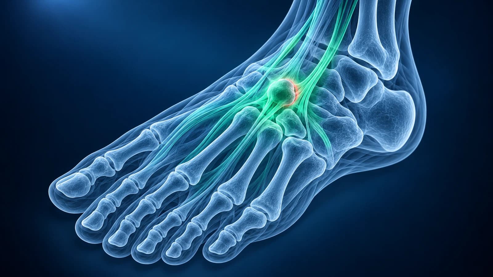
A soft, rounded lump on top of the foot is most often a ganglion cyst, and a hard bump over the middle of the foot is usually an arthritis spur. Pain along a tendon under tight laces suggests extensor tendinitis, and pain after a twist with bruising on the sole needs a Lisfranc check. Ultrasound sorts out lumps; a standing X-ray sorts out bones.
Most lumps on the foot are harmless, but a few need a faster look. This hub explains the common causes on the top of the foot and the firm lump in the arch, which test is used for each and which signs mean a lump should be checked soon. It is educational and is not a diagnosis.
Which foot lumps should be checked soon?
UK cancer referral guidance advises an urgent ultrasound for an unexplained lump that keeps getting bigger. Ask for a prompt review if a lump on the foot:
- is growing over weeks to months, or has appeared where a lump was removed before
- is 5 cm or more across, feels hard and fixed, or sits deep rather than just under the skin
- is painful, especially at rest or at night, or the skin over it changes colour or breaks down
- comes with fever, a hot red foot or, in diabetes, any wound nearby
Which test shows which cause of top-of-foot pain or a lump?
The ACR Appropriateness Criteria for soft-tissue masses rate an X-ray or an ultrasound as the first test for a lump just under the skin, an X-ray first for a deeper lump, and MRI without and with contrast when those first tests do not explain it. For midfoot pain without a lump, the chronic foot pain criteria also start with radiographs.
| Cause | What you notice | Test that shows it best | Typical report wording |
|---|---|---|---|
| Ganglion cyst | Soft, smooth lump that may change size | Ultrasound | Anechoic, lobulated cystic lesion arising from a joint or tendon sheath |
| Midfoot arthritis spur | Hard bump over the middle of the foot, aching when walking | Standing X-ray, side view | Dorsal osteophyte, joint space narrowing at the tarsometatarsal joints |
| Lisfranc injury | Midfoot pain after a twist, bruising on the sole | Weight-bearing X-ray; CT or MRI if normal | Widening between the 1st and 2nd metatarsal bases, fleck sign |
| Extensor tendinitis | Pain along a tendon on top of the foot, worse with tight laces | Examination; ultrasound if unclear | Extensor tenosynovitis, fluid in the tendon sheath |
| Plantar fibroma (sole) | Firm, rubbery nodule in the arch | Ultrasound; MRI for large or multiple nodules | Fusiform hypoechoic nodule within the plantar fascia |
Where is the lump or pain on top of your foot?
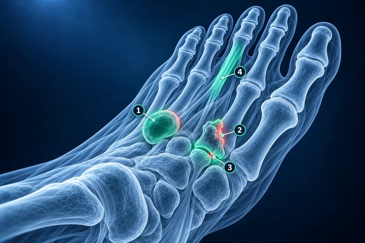
A lump you can move slightly and that feels like a firm water balloon is usually a cyst. A lump that feels like bone and does not move is usually a spur. Tenderness in a line running toward the toes follows a tendon, while tenderness across the width of the midfoot follows the Lisfranc joints.
Is the soft lump on top of my foot a ganglion cyst?
A ganglion cyst is a pocket of thick, jelly-like fluid that grows out of a joint capsule or tendon sheath. It may come and go and often hurts only when a shoe presses on it. On ultrasound it looks black, with a thin wall and sometimes a narrow neck leading back to the joint; that appearance confirms it without a needle. MRI is used when the lump is deep, does not look like a simple cyst or is pressing on a nerve.
What does a hard bump over the midfoot mean?
A bony ridge on top of the midfoot, often over the joints at the base of the second and third metatarsals, is usually an osteophyte from midfoot arthritis. The side-view standing X-ray shows the spur and the narrowed joint, and the front view checks alignment. The spur itself can irritate the nerve that runs over it, causing tingling toward the toes. Sometimes the same bump follows an old Lisfranc injury that has worn the joint.
Could top-of-foot pain after a twist be a Lisfranc injury?
Yes, and it is one not to miss. A Lisfranc injury damages the ligaments that hold the midfoot bones together. Bruising on the sole, pain when standing or pushing off and swelling across the midfoot are warning signs. Subtle injuries can look normal on a non-weight-bearing X-ray, which is why standing views, comparison with the other foot, CT or MRI are added; see Lisfranc injury missed on X-ray. You can check the alignment of your own images in our free X-ray viewer.
What is extensor tendinitis on top of the foot?
The tendons that lift the toes and the foot run over the top of the midfoot just under the laces. Tight lacing, a new shoe, uphill running or a high arch can inflame them. The pain follows the line of the tendon and increases when you pull the toes up against resistance. X-rays are normal; ultrasound can show fluid around the tendon if the diagnosis is in doubt. Changing the lacing pattern to skip the sore eyelets often helps quickly.
What is the firm lump in the arch of my foot?
A firm, rubbery nodule in the middle of the arch that moves with the fascia is usually a plantar fibroma, a benign thickening of the plantar fascia that can be single or multiple. Ultrasound shows a spindle-shaped dark nodule inside the fascia. MRI is used for larger or multiple nodules and before any surgery, because removal can be followed by regrowth. A fibroma is not the same as plantar fasciitis, which thickens the fascia at the heel rather than forming a lump in the arch.
Can gout cause a lump on the foot?
Yes. Long-standing gout can leave chalky deposits called tophi over joints, including the midfoot and the big toe. They feel hard and may show white material through the skin. Ultrasound can show a characteristic double contour over the cartilage, and dual-energy CT can map urate deposits. More on gout in the foot.
Should I have an ultrasound or an MRI for a foot lump?
Ultrasound first for most small lumps near the surface: it shows whether the lump is fluid or solid, where it comes from and whether it has blood flow. MRI, usually with contrast, is the next step when a lump is solid and unexplained, deep, growing or larger than expected, and before surgery. MRI images open privately in our free MRI viewer, and foot MRI vs X-ray vs ultrasound compares the three tests in more detail.
Frequently Asked Questions
What is the most common lump on top of the foot?
A ganglion cyst is the most common soft lump, and a bony spur from midfoot arthritis is the most common hard bump. Ultrasound tells a cyst from a solid lump, and an X-ray shows a spur.
Why does the top of my foot hurt with no injury?
Common causes are extensor tendinitis from tight laces or new shoes, midfoot arthritis and stress fractures of the metatarsals or the navicular. If the pain persists or there is swelling, an X-ray is the usual first test.
Can a foot lump be cancer?
It is rare. Most foot lumps are cysts, fibromas or spurs. A lump that is growing, deep, hard, painful at rest or returning after removal should be imaged promptly, usually starting with ultrasound and then MRI.
Does a ganglion cyst on the foot need to be removed?
Not always. Many are left alone or managed with shoe changes, and some shrink by themselves. Aspiration or surgery is considered when the cyst hurts, presses on a nerve or makes shoes impossible, and it can come back.
Can AI read my foot lump ultrasound report?
AI can explain terms such as anechoic, hypoechoic or vascularity and what they usually mean for a lump. Whether the lump needs a biopsy or removal is decided by your clinician.
Sources
- Expert Panel on Musculoskeletal Imaging; Garner HW, Wessell DE, Lenchik L, et al. ACR Appropriateness Criteria Soft Tissue Masses: 2022 Update. J Am Coll Radiol. 2023;20(5S):S234-S245.
- Expert Panel on Musculoskeletal Imaging; Said N, Fox MG, Nacey NC, et al. ACR Appropriateness Criteria Chronic Foot Pain: Update 2025. J Am Coll Radiol. 2026;23(4):681-699.
- National Institute for Health and Care Excellence. Suspected cancer: recognition and referral (NG12).
- NHS Scotland Centre for Sustainable Delivery. Scottish Referral Guidelines for Suspected Cancer: Sarcoma and Bone Cancers. Right Decision Service.
- Grimer R, Judson I, Peake D, Seddon B. Guidelines for the management of soft tissue sarcomas. Sarcoma. 2010;2010:506182.
- American Academy of Orthopaedic Surgeons. Lisfranc (Midfoot) Injury. OrthoInfo.
- Stern JM, Bergman R, Singh S. Lisfranc Dislocation. In: StatPearls [Internet]. Treasure Island (FL): StatPearls Publishing.
- Neogi T, Jansen TL, Dalbeth N, et al. 2015 Gout classification criteria: an American College of Rheumatology/European League Against Rheumatism collaborative initiative. Arthritis Rheumatol. 2015;67(10):2557-2568.
Related Articles
Why Lisfranc midfoot injuries are missed on first X-rays, the alignment lines and fleck sign to check, and when standing X-rays, CT or MRI are needed.
X-ray, MRI, ultrasound or CT for foot pain? What each test shows and misses, which one usually comes first, and how the choice changes for stress fractures, neuromas, plantar fasciitis, Lisfranc injuries and the diabetic foot.
Ball of foot pain by cause: metatarsalgia, Morton's neuroma, plantar plate tear, stress fracture, Freiberg disease and sesamoiditis, and the test for each.
Related Conditions
Understand your imaging before your next appointment
MRI, X-ray or CT from a CD, folder or ZIP. A PDF report or photos of the films work too.
Free to open and preview, no account needed. An optional AI report from $10 explains your images in plain language, with questions for your doctor.
Your raw files stay on this device. Only rendered images and minimal context are sent when you request an AI report.
Medical Disclaimer: This page is for informational and educational purposes only. It does not constitute medical advice, diagnosis, or treatment. AI-generated analysis may contain errors. Always consult a qualified healthcare professional for medical decisions. Full Disclaimer