
Plantar Fibroma: What a Lump in the Arch Looks Like on Imaging
A plantar fibroma is a firm, benign nodule that grows inside the plantar fascia, the tough band under the arch. Ultrasound is usually the first and often the only test needed: it shows an oval nodule within the fascia. MRI is added when the lump looks unusual, is large or deep, or before surgery. A lump that grows quickly needs prompt assessment.
Common Symptoms
- A firm, rubbery lump in the middle of the arch that you can feel through the skin of the sole
- A lump that moves with the band under the arch rather than sliding freely under the skin
- Discomfort when the lump is pressed by a shoe, an arch support or a hard floor
- Several small nodules along the arch, sometimes in both feet
- Similar cords or nodules in the palm of the hand that pull the fingers into a bent position
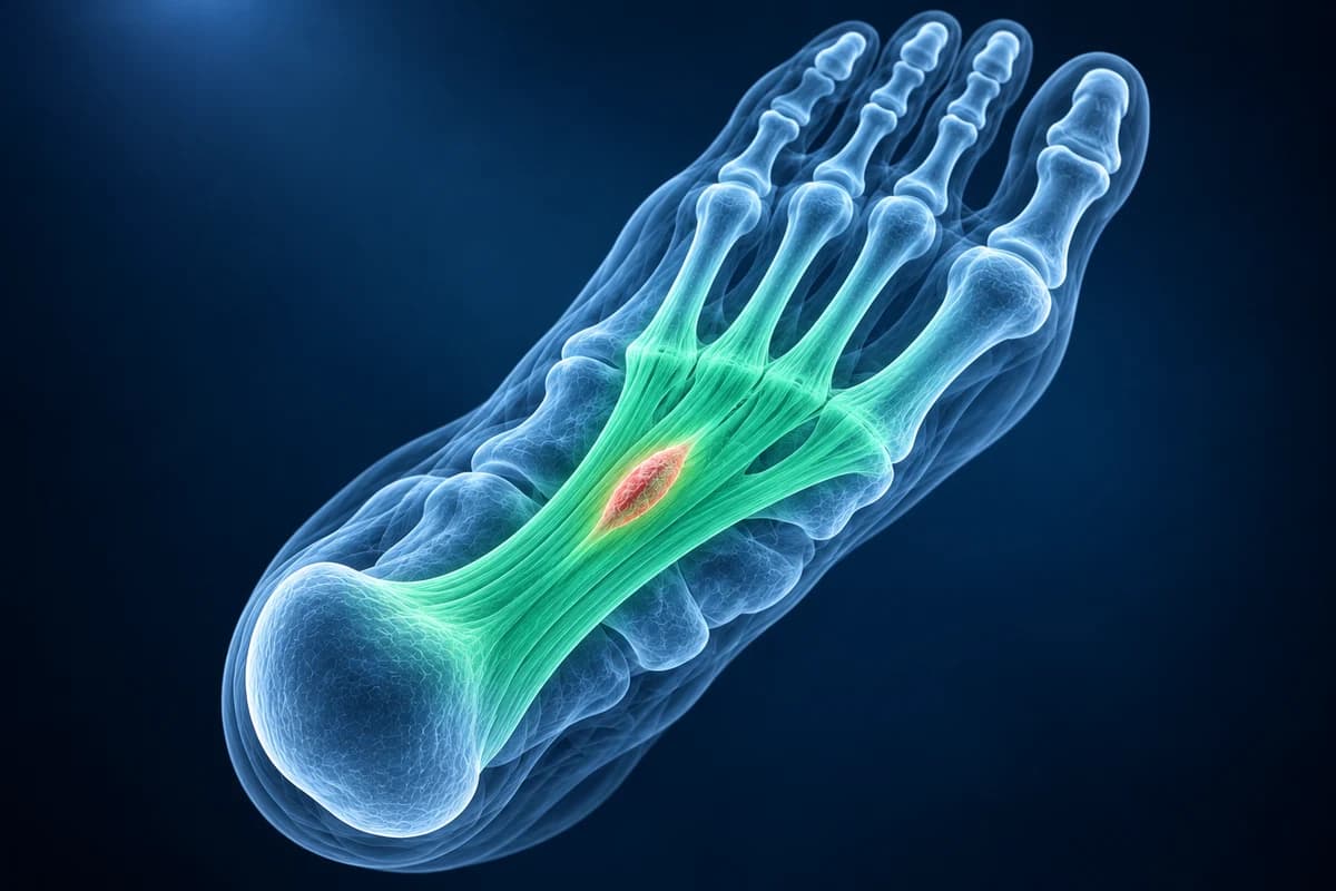
Plantar fibromas belong to a family of conditions called fibromatoses, in which fibrous tissue overgrows within a fascia. They grow slowly, do not spread and are often painless until shoes press on them. The condition is also called plantar fibromatosis or Ledderhose disease, and it can produce one nodule or several, in about a quarter of people in both feet. This page explains how the lump is confirmed, the phrases your report may use and the warning signs that a lump needs faster assessment. It is educational guidance, not a diagnosis.
Is a lump in the arch of my foot serious? Red flags
A firm lump that sits within the band under the arch is usually a plantar fibroma. Soft-tissue cancers called sarcomas are rare, but they can occur in the foot and can look harmless at first. UK sarcoma guidelines list features that should prompt urgent assessment, and NICE recommends considering an urgent ultrasound, within two weeks, for an unexplained lump that is getting bigger. Ask for prompt review if the lump:
- is growing steadily over weeks or months, rather than staying the same size;
- is larger than 5 cm across;
- lies deep, rather than within the band just under the skin;
- is painful, although many harmless fibromas also hurt when pressed;
- was thought to be harmless but has started to grow or change.
These signs do not mean cancer; they mean the lump should be imaged and reviewed quickly, sometimes at a specialist sarcoma center, before anyone cuts it out.
Which test confirms a plantar fibroma?
Ultrasound is the first test for a lump just under the skin. The American College of Radiology Appropriateness Criteria for soft-tissue masses support ultrasound for superficial lumps, and a plantar fibroma has a recognizable look: a smooth, spindle-shaped, darker nodule sitting within the fascia, usually toward the inner side of the arch, with little or no blood flow on Doppler. The scan takes minutes, uses no radiation and can compare both feet.
An X-ray of the foot is usually normal with a fibroma. It is still useful when the lump is hard, to check for bone involvement or calcification, which a fibroma does not usually show.
MRI with contrast is added when ultrasound does not give a clear answer, when the lump is large, deep or growing, or before surgery to map how far the tissue extends. Fibromas typically appear dark on both T1 and T2 images because they are dense fibrous tissue, and they enhance to a variable degree after contrast. When imaging is not typical, a biopsy planned by a specialist team settles the diagnosis. MRI files can be opened in the free MRI viewer, and the comparison of foot MRI, X-ray and ultrasound explains what each test can see.
What does “nodule within the plantar fascia” mean on your report?
These are the phrases arch ultrasound and MRI reports use for a plantar fibroma. The foot MRI report phrases guide covers the wider vocabulary.
- “Fusiform hypoechoic nodule within the medial cord of the plantar fascia”: a spindle-shaped, darker lump inside the inner part of the band, the classic ultrasound look of a fibroma. Ask whether any feature looks atypical.
- “No significant internal vascularity”: Doppler shows little blood flow in the lump, which fits a benign fibrous nodule. Ask whether the size should be checked again later.
- “Low signal on T1- and T2-weighted images” on MRI: the lump is dense fibrous tissue rather than fluid or fat. Ask whether the MRI appearance is typical enough to avoid a biopsy.
- “Multiple nodules, consistent with plantar fibromatosis (Ledderhose disease)”: more than one nodule, sometimes in both feet. Ask whether the palms should be checked as well.
- “Indeterminate soft-tissue mass; further evaluation recommended”: the radiologist cannot call the lump a typical fibroma. Ask whether you should be referred to a sarcoma service before any surgery.
What We Look For on Arch Lump Imaging
- Whether the lump lies inside the plantar fascia or deeper among the muscles of the sole
- Nodule size, shape and edges, and the number of nodules in each foot
- Internal blood flow on Doppler ultrasound and enhancement after MRI contrast
- Signal on T1 and T2 MRI images compared with fibrous tissue, fluid and fat
- Involvement of nearby nerves, tendons, the skin or bone
- Features that suggest another kind of lump, such as a cyst, a nerve tumor or a foreign-body reaction
Is it a plantar fibroma or plantar fasciitis?
Both involve the same band, but they behave differently. Plantar fasciitis causes pain where the fascia attaches under the heel, worst with the first morning steps, and imaging shows a thickened band at the heel without a separate lump. A fibroma is a distinct lump further forward, in the middle of the arch, and is often painless unless pressed.
Other lumps in the foot follow different patterns. A soft, fluid-filled lump that changes size, usually on the top of the foot, is more often a ganglion cyst. Pain and tingling between two toes in the forefoot points toward a Morton's neuroma. The guide to foot pain and lumps walks through the common causes and the right first test for each.
How is a plantar fibroma treated?
A fibroma that does not hurt can simply be watched. For a painful one, the first steps take pressure off the lump: soft insoles with a cut-out or a recess under the nodule, cushioned shoes and avoiding rigid arch supports that push into it. Stretching of the calf and fascia helps some people. Steroid injections can shrink a painful nodule for a while, and some centers offer radiotherapy for painful Ledderhose disease, which eased pain more than a sham treatment in a randomised trial.
Surgery is kept for nodules that stay painful or keep growing. Fibromas tend to return after simple removal of the lump, so surgeons sometimes remove a wider part of the fascia, which lowers the chance of recurrence but brings its own risks of a painful scar, nerve injury and flattening of the arch. A frank discussion of these trade-offs with a foot and ankle surgeon matters more than the scan alone.
Frequently Asked Questions
Is a plantar fibroma cancer?
No. A plantar fibroma is benign and does not spread to other parts of the body. It can grow slowly and come back after surgery. Because rare cancers can also form lumps in the foot, a lump that grows quickly, is large, deep or painful should be imaged and reviewed promptly.
Can an ultrasound diagnose a plantar fibroma?
In most cases, yes. A spindle-shaped, darker nodule within the plantar fascia with little blood flow is a typical picture, and together with a typical examination it is often enough. MRI or a biopsy is added when the appearance is unusual.
What is Ledderhose disease?
Ledderhose disease is another name for plantar fibromatosis, which can mean a single nodule or several, sometimes in both feet. It is related to Dupuytren's disease in the palm, and some people have both. It is benign but tends to recur after surgery.
Should a plantar fibroma be removed?
Usually not at first. Most are managed with padding, shoe changes and sometimes injections. Surgery is considered when the lump stays painful or grows, and the choice between removing the nodule and removing more of the fascia depends on the size, the number of nodules and the recurrence risk.
Can AI read my foot ultrasound or MRI report for a lump?
A nodule within the plantar fascia may be a fibroma, but a growing or atypical lump needs clinical review. AI cannot establish that a lump is benign.
Sources
- Murphey MD, Ruble CM, Tyszko SM, Zbojniewicz AM, Potter BK, Miettinen M. From the archives of the AFIP: musculoskeletal fibromatoses: radiologic-pathologic correlation. Radiographics. 2009;29(7):2143-2173.
- Expert Panel on Musculoskeletal Imaging; Garner HW, Wessell DE, Lenchik L, et al. ACR Appropriateness Criteria Soft Tissue Masses: 2022 Update. J Am Coll Radiol. 2023;20(5S):S234-S245.
- Dangoor A, Seddon B, Gerrand C, Grimer R, Whelan J, Judson I. UK guidelines for the management of soft tissue sarcomas. Clin Sarcoma Res. 2016;6:20.
- National Institute for Health and Care Excellence. Suspected cancer: recognition and referral (NG12).
- Young JR, Sternbach S, Willinger M, Hutchinson ID, Rosenbaum AJ. The etiology, evaluation, and management of plantar fibromatosis. Orthop Res Rev. 2019;11:1-7.
- de Haan A, van Nes JGH, Kolff MW, et al. Radiotherapy for Ledderhose disease: results of the LedRad-study, a prospective multicentre randomised double-blind phase 3 trial. Radiother Oncol. 2023;185:109718.
Understand your imaging before your next appointment
MRI, X-ray or CT from a CD, folder or ZIP. A PDF report or photos of the films work too.
Free to open and preview, no account needed. An optional AI report from $10 explains your images in plain language, with questions for your doctor.
Your raw files stay on this device. Only rendered images and minimal context are sent when you request an AI report.
Medical Disclaimer: This page is for informational and educational purposes only. It does not constitute medical advice, diagnosis, or treatment. AI-generated analysis may contain errors. Always consult a qualified healthcare professional for medical decisions. Full Disclaimer