
Ganglion Cyst of the Foot: How Imaging Confirms It and What Comes Next
A ganglion cyst on the foot is a smooth sac of thick, jelly-like fluid, most often on the top of the midfoot or around the ankle, attached to a joint or a tendon sheath. Ultrasound usually confirms it within minutes by showing a fluid-filled lump with no blood flow inside. Many cysts are harmless; aspiration and surgery help, but cysts often return.
Common Symptoms
- A smooth, rounded swelling on the top of the foot or around the ankle that may feel firm or squashy
- A lump that grows, shrinks or disappears for a while, often getting bigger with activity
- Soreness where the lump rubs against shoe laces, straps or the edge of a shoe
- Tingling, burning or numbness spreading toward the toes when the lump presses on a nerve
- An ache in the joint or tendon underneath, especially after long walks
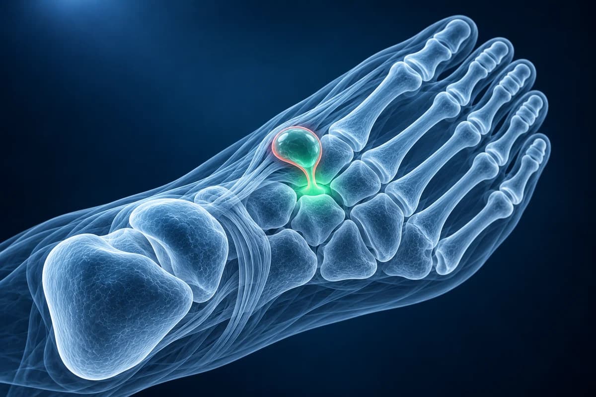
A ganglion forms when joint or tendon fluid leaks through a weak spot in the lining and collects in a pouch with a thin wall. The pouch keeps a narrow stalk back to its source, which is why it can swell with activity and shrink with rest. Foot ganglia are especially bothersome because shoes press right on them. This page explains how imaging tells a ganglion from other lumps, the report phrases you may see and how treatment choices compare. It is educational guidance, not a diagnosis.
Where do ganglion cysts form on the foot?
In a surgical series of foot and ankle ganglia, about two-thirds sat on the top of the foot and ankle, and more grew from tendon sheaths than from joints. Others form near the tarsal tunnel behind the inner ankle bone or, less often, on the sole or under the big toe.
A ganglion on top of the foot sometimes grows from a joint already worn by midfoot arthritis, in which case a bone spur and a cyst can sit side by side. The deep peroneal nerve runs across the same area, so a cyst there can cause tingling in the web space between the big toe and the second toe.
Is it a ganglion or something else? Warning signs in a foot lump
A ganglion typically feels smooth and rubbery, moves slightly, changes size over time and may glow when a torch is held against it. A lump that does not fit this pattern should be scanned rather than assumed to be a cyst. UK sarcoma guidance and NICE referral advice treat these features as reasons for urgent imaging and specialist review:
- steady growth rather than the up-and-down size changes typical of a ganglion;
- a diameter of more than 5 cm;
- a lump that lies deep rather than just under the skin;
- pain in the lump that is not explained by shoe pressure;
- a lump thought to be harmless that starts to grow or change.
A red, hot, tender swelling with fever suggests infection and needs same-day care, especially if you have diabetes. A chalky lump near the big toe in someone with gout may be a tophus rather than a cyst.
Do you need an ultrasound or MRI for a ganglion cyst on the foot?
Ultrasound is the usual first test. The American College of Radiology Appropriateness Criteria for soft-tissue masses support ultrasound for lumps near the surface, and a ganglion has a distinctive look: a dark, fluid-filled, well-defined pocket, often with several lobes, bright tissue behind it where sound passes easily through fluid, and no blood flow on Doppler. Often the scan also traces the stalk back to the joint or tendon it comes from.
An X-ray adds information when the lump feels hard or sits over a painful joint, because it shows arthritis, spurs or an underlying bone problem that ultrasound cannot see well.
MRI is added when the lump is deep, unusual on ultrasound, causing nerve symptoms without an obvious swelling, or before surgery to map the stalk. On MRI a ganglion is bright on fluid-sensitive images, and after contrast only its thin wall lights up. MRI files can be opened in the free MRI viewer, and the guide to top of foot pain and lumps compares ganglia with the other common lumps and bumps.
What does “anechoic lobulated cystic lesion” mean on your report?
These are the phrases foot ultrasound and MRI reports use for a ganglion. The foot MRI report phrases guide explains the wider vocabulary.
- “Anechoic, lobulated cystic lesion with posterior acoustic enhancement”: a fluid-filled lump with several rounded pockets, the classic ultrasound look of a ganglion. Ask whether anything about it looks atypical.
- “No internal vascularity on color Doppler”: no blood flow inside, which supports a simple cyst rather than a solid lump. Ask whether a follow-up scan is needed.
- “Communicating neck with the second tarsometatarsal joint” or “arising from the extensor tendon sheath”: the stalk leads back to its source. Ask how this affects the chance of return after aspiration or surgery.
- “Thin peripheral rim enhancement” on MRI: after contrast only the wall lights up, as expected for a cyst. Ask whether the MRI rules out a solid component.
- “Complex cystic lesion with solid components; further evaluation recommended”: the lump is not a simple ganglion. Ask what the next step is, which may be MRI with contrast or a specialist review.
What We Look For on Foot Lump Imaging
- Whether the lump is fluid-filled or solid, and the number of lobes or internal walls
- Blood flow inside the lump on Doppler and enhancement pattern on MRI
- The stalk and its source: a midfoot joint, the ankle joint or a tendon sheath
- Pressure on nearby nerves, especially the deep peroneal nerve on the top of the foot
- Arthritis, bone spurs or bone changes in the joint beneath the cyst
- Size and depth, compared with any earlier scans
Should a foot ganglion be drained or removed, and will it come back?
A ganglion that causes no pain or shoe trouble can be left alone, and some shrink or disappear on their own. Padding, wider shoes and lacing that skips the eyelets over the lump are simple first steps. Hitting the cyst with a heavy object, an old home remedy, can injure the foot and is not recommended.
Aspiration drains the fluid through a needle, ideally under ultrasound guidance, sometimes with a steroid injection. It is quick and relieves pressure, but because the wall and stalk stay behind, the cyst often fills again. Surgical excision removes the sac together with its stalk and has a lower recurrence rate in published foot series, though recurrences still happen and the scar, nearby nerves and tendons carry their own risks. Your surgeon may want an MRI first to find the exact source.
A firm lump in the middle of the arch on the sole of the foot is more often a plantar fibroma than a ganglion, and is treated differently.
Frequently Asked Questions
What does a ganglion cyst on the top of the foot look like on ultrasound?
It appears as a dark, well-defined pocket of fluid, often with a few rounded lobes and bright tissue behind it, and no blood flow inside on Doppler. The scan often shows a thin stalk leading to the joint or tendon sheath underneath.
Can a ganglion cyst on the foot go away on its own?
Sometimes. Ganglia can shrink or vanish without treatment and can also return later. If the cyst is not painful and does not press on a nerve, watching it is a reasonable choice, provided it has been confirmed as a ganglion.
Why did my ganglion come back after it was drained?
Draining empties the fluid but leaves the wall and the stalk that connects to the joint or tendon sheath, so fluid can collect again. Surgery that removes the stalk lowers the chance of return, but no treatment guarantees a cyst will never come back.
When should I worry about a lump on my foot?
Get it checked promptly if it keeps growing, is larger than 5 cm, lies deep, hurts without shoe pressure or starts to change after being called harmless. Same-day care is needed for a hot, red, swollen lump with fever. Most foot lumps are benign, but these signs mean it should be imaged.
Can AI read my foot ultrasound report for a cyst?
Ultrasound can show a fluid-filled cyst, but atypical or growing lumps need clinical review. AI cannot confirm a lump is benign or decide whether it needs biopsy.
Sources
- Rozbruch SR, Chang V, Bohne WH, Deland JT. Ganglion cysts of the lower extremity: an analysis of 54 cases and review of the literature. Orthopedics. 1998;21(2):141-148.
- Pontious J, Good J, Maxian SH. Ganglions of the foot and ankle. A retrospective analysis of 63 procedures. J Am Podiatr Med Assoc. 1999;89(4):163-168.
- Ahn JH, Choy WS, Kim HY. Operative treatment for ganglion cysts of the foot and ankle. J Foot Ankle Surg. 2010;49(5):442-445.
- Expert Panel on Musculoskeletal Imaging; Garner HW, Wessell DE, Lenchik L, et al. ACR Appropriateness Criteria Soft Tissue Masses: 2022 Update. J Am Coll Radiol. 2023;20(5S):S234-S245.
- Dangoor A, Seddon B, Gerrand C, Grimer R, Whelan J, Judson I. UK guidelines for the management of soft tissue sarcomas. Clin Sarcoma Res. 2016;6:20.
- National Institute for Health and Care Excellence. Suspected cancer: recognition and referral (NG12).
Understand your imaging before your next appointment
MRI, X-ray or CT from a CD, folder or ZIP. A PDF report or photos of the films work too.
Free to open and preview, no account needed. An optional AI report from $10 explains your images in plain language, with questions for your doctor.
Your raw files stay on this device. Only rendered images and minimal context are sent when you request an AI report.
Medical Disclaimer: This page is for informational and educational purposes only. It does not constitute medical advice, diagnosis, or treatment. AI-generated analysis may contain errors. Always consult a qualified healthcare professional for medical decisions. Full Disclaimer