
Foot X-ray Angles Explained: Bunion, Arch and Heel Measurements
What the numbers on your foot X-ray mean: hallux valgus angle, intermetatarsal angle, Meary's angle, calcaneal pitch and Böhler's angle, with normal ranges and how each is measured.
Key Takeaways
- HVA above 15° or IMA above 9° on a standing X-ray is the usual radiographic definition of a bunion
- Meary’s angle and calcaneal pitch describe the arch on a standing side view
- A flattened Böhler’s angle supports a calcaneal fracture that has crushed the joint surface
- Lisfranc injuries are judged by alignment and a widened gap, not by an angle
- Angles guide the choice of treatment; symptoms decide whether treatment is needed
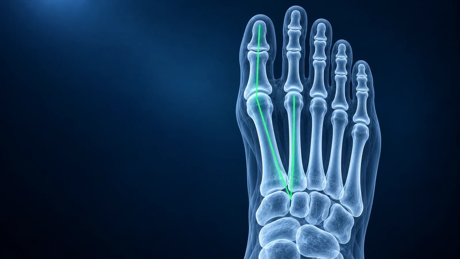
Foot X-ray angles turn a picture into numbers that can be compared over time. The two most common are the hallux valgus angle (HVA) and the intermetatarsal angle (IMA): an HVA above 15° or an IMA above 9° on a standing X-ray is generally considered abnormal and is the radiographic definition of a bunion. Other angles describe the height of the arch, the shape of the heel bone and the alignment of the midfoot.
Normal ranges vary slightly between references, measurement errors of a few degrees are common even between experts, and the number alone never decides treatment. Use this page to understand your report, then discuss the values alongside your symptoms with your clinician.
What is a normal hallux valgus angle?
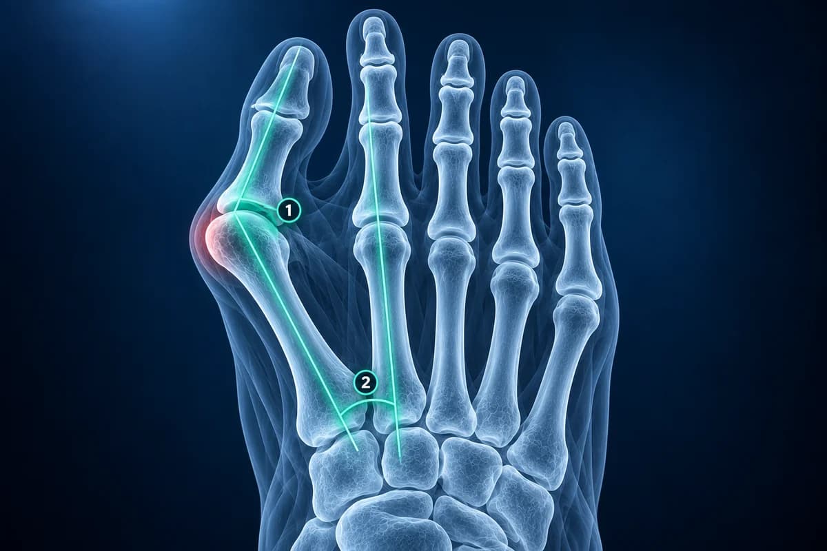
The hallux valgus angle measures how far the big toe leans toward the second toe compared with its own metatarsal. Below 15° is considered normal. As the angle grows, the metatarsal head pushes out at the side of the foot and the bump of a bunion becomes visible. A widely used grading, combining HVA and IMA, is:
- Normal: HVA below 15° and IMA below 9°
- Mild: HVA 15°–20°, IMA 9°–11°
- Moderate: HVA 20°–40°, IMA 11°–16°
- Severe: HVA above 40°, IMA above 16°
Surgeons use these grades mainly to choose between operations, not to decide whether surgery is needed at all; pain, shoe problems and function drive that decision. Our bunion surgery X-ray before and after guide shows how the same angles are used to judge correction.
What does the intermetatarsal angle tell you?
The IMA measures how far the first metatarsal has splayed away from the second. Normal is below 9°. A large IMA means the deformity starts further back in the foot, at the base of the first metatarsal, so a simple shave of the bump would not straighten it. Reports may add related terms:
- “Lateral sesamoid displacement”: the two small bones under the big toe joint have slid outward from their groove, a sign that the deformity has progressed.
- “Distal metatarsal articular angle (DMAA)”: the tilt of the joint surface on the metatarsal head itself, which some surgeons factor into the choice of correction.
- “Hallux valgus interphalangeus”: an extra lean within the big toe, between its two bones, rather than at the bunion joint.
- “Joint congruent / incongruent”: whether the toe still sits squarely on the metatarsal head or has started to slide off it.
Which X-ray angles measure a flat foot or high arch?
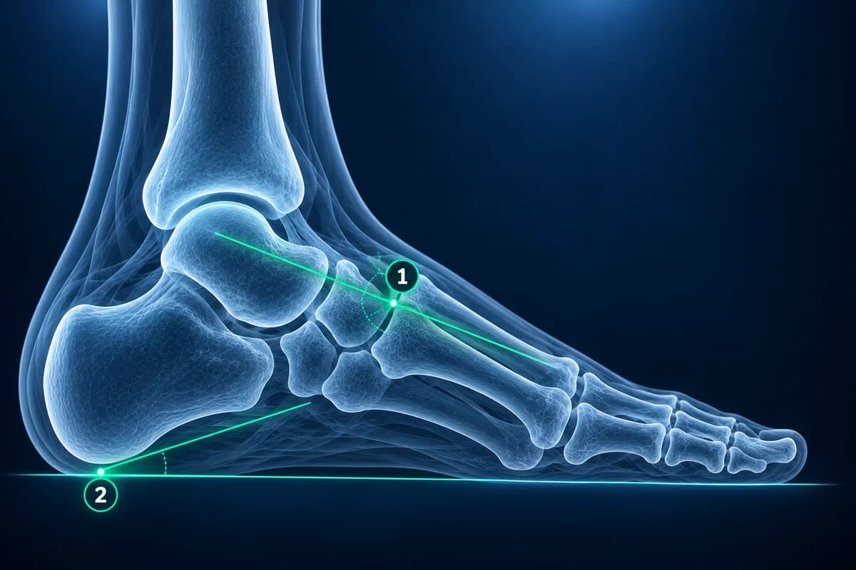
Arch angles are measured on a standing side (lateral) view. Meary’s angle, also called the lateral talus to first metatarsal angle, compares a line through the talus with a line along the first metatarsal. In a well-aligned foot the two lines are nearly continuous, close to 0°. When the line breaks downward at the arch, it indicates flat foot; when it breaks upward, it indicates a high arch (cavus foot). The calcaneal pitch is the angle between the underside of the heel bone and the floor; it is lower in flat feet and higher in high-arched feet.
On the top-down view, reports may mention “talonavicular uncoverage”, meaning the head of the talus is no longer fully capped by the navicular because the forefoot has swung outward, and the AP talus to first metatarsal angle, which captures the same outward drift. In a study comparing adults with painful flat feet and controls on standing films, the lateral talus to first metatarsal angle was the clearest separator (about 21° on average in the flat feet versus about 7° in the controls), and talar head uncoverage was the most telling measurement on the top-down view. In a child or teenager with a stiff flat foot, the side view is also checked for the “C-sign” or “anteater nose sign” of a tarsal coalition.
What is Böhler’s angle on a heel X-ray?
Böhler’s angle is drawn on the side view of the heel bone from two lines that meet at the top of its joint surface. Textbooks often quote a normal range of about 20° to 40°; a study of 128 uninjured British heel X-rays found an average of about 36° with a wide spread, so comparison with the other heel helps when the value is borderline. When a fall from height crushes the joint surface, the angle flattens or even reverses, which helps confirm a calcaneal fracture and track how well it was restored after surgery. The related crucial angle of Gissane, measured on the same view, is often reported alongside it. A CT scan is usually added to map a heel fracture in detail.
What measurements are used for a Lisfranc injury?
Instead of an angle, a midfoot injury is judged by alignment and gaps. The inner edge of the second metatarsal should line up with the inner edge of the middle cuneiform. A gap of more than about 2 mm between the bases of the first and second metatarsals, especially on a standing film or compared with the other foot, suggests the ligament that binds them is torn. A small “fleck” of bone in that gap supports the diagnosis. Read more on the Lisfranc injury page and in why a Lisfranc injury can be missed on X-ray.
Why do two reports give different angles for the same foot?
Small differences are expected. The reader chooses where to place each midpoint, the foot may be rotated slightly differently on the second film, and a seated X-ray gives lower numbers than a standing one. A change of a few degrees between two visits is usually within measurement error; a steady rise over several standing films is more meaningful. If you want to see the lines for yourself, open your images in the free X-ray viewer, which keeps the files in your browser. Our guide on how to read a foot X-ray covers the rest of the film.
Which imaging test comes before measuring foot angles?
The ACR recommends radiographs first for unexplained chronic foot pain in adults. Further imaging depends on the suspected bone, joint or soft-tissue cause, rather than an angle measurement alone.
Frequently Asked Questions
What hallux valgus angle needs surgery?
No angle on its own means surgery is needed. Surgery is considered when a bunion causes ongoing pain or footwear problems despite non-surgical care. The HVA and IMA then help the surgeon choose which procedure fits the deformity.
Can my bunion angle be measured on a non-weight-bearing X-ray?
It can be measured, but it usually underestimates the deformity. Bunion grading is designed for standing films, so ask for a weight-bearing view if the angles will guide treatment.
Does a low calcaneal pitch mean I have flat feet?
A low calcaneal pitch is one feature of a flat foot, but the diagnosis also depends on Meary’s angle, the view from above and the clinical exam. Many flexible flat feet cause no symptoms and need no treatment.
Can AI measure foot X-ray angles?
Research tools can measure angles such as the HVA automatically, and AI can explain the numbers in your report. Treat any automated value as an estimate and confirm it with the radiologist or surgeon who will plan your care.
Sources
- Coughlin MJ, Jones CP. Hallux valgus: demographics, etiology, and radiographic assessment. Foot Ankle Int. 2007;28(7):759-777.
- Robinson AH, Limbers JP. Modern concepts in the treatment of hallux valgus. J Bone Joint Surg Br. 2005;87(8):1038-1045.
- Younger AS, Sawatzky B, Dryden P. Radiographic assessment of adult flatfoot. Foot Ankle Int. 2005;26(10):820-825.
- Willmott H, Stanton J, Southgate C. Böhler’s angle: what is normal in the uninjured British population? Foot Ankle Surg. 2012;18(3):187-189.
- Kalia V, Fishman EK, Carrino JA, et al. Epidemiology, imaging, and treatment of Lisfranc fracture-dislocations revisited. Skeletal Radiol. 2012;41(2):129-136.
Related Articles
A plain-language guide to your foot X-ray: the three standard views, how to trace alignment from heel to toes, and what fractures, fleck signs, accessory bones and arthritis look like.
What a bunion X-ray measures before surgery, what a normal post-op X-ray looks like, how long the bone cut takes to heal, and which report findings need attention.
X-ray, MRI, ultrasound or CT for foot pain? What each test shows and misses, which one usually comes first, and how the choice changes for stress fractures, neuromas, plantar fasciitis, Lisfranc injuries and the diabetic foot.
Related Conditions
Understand your imaging before your next appointment
MRI, X-ray or CT from a CD, folder or ZIP. A PDF report or photos of the films work too.
Free to open and preview, no account needed. An optional AI report from $10 explains your images in plain language, with questions for your doctor.
Your raw files stay on this device. Only rendered images and minimal context are sent when you request an AI report.
Medical Disclaimer: This page is for informational and educational purposes only. It does not constitute medical advice, diagnosis, or treatment. AI-generated analysis may contain errors. Always consult a qualified healthcare professional for medical decisions. Full Disclaimer