
Plantar Fasciitis Recovery Timeline: Month by Month
How long plantar fasciitis lasts, what changes month by month, why morning pain is the last to go, and when imaging or a different diagnosis should be considered.
Key Takeaways
- Most plantar fasciitis improves within six to twelve months of consistent non-surgical care
- First-step morning pain is the last symptom to settle and the best one to track
- Imaging is not needed at first; it mainly rules out stress fracture, tear or other causes
- A fascia thicker than about 4 mm on ultrasound supports the diagnosis and thins slowly
- Heel spurs are common in people without pain and are not the target of treatment
Most people with plantar fasciitis are clearly better within six to twelve months of consistent non-surgical care, and many notice the first real change after six to eight weeks. The sharp first-step pain in the morning is usually the last symptom to go, and a plantar fascia thicker than 4 mm on ultrasound can stay thick after the pain has settled.
That slow arc is frustrating, because the heel often feels a little better for a week and then flares after a long walk or a new pair of shoes. Knowing what a normal course looks like makes it easier to stick with the boring parts of treatment and to recognise when the pattern is not normal. This page is general education; it cannot tell you whether your heel pain is plantar fasciitis or something else, which is a clinical question.
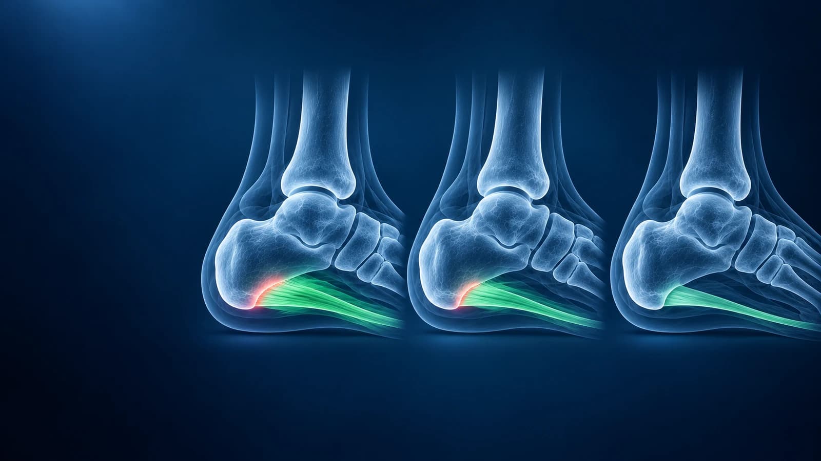
How long does plantar fasciitis last?
Plantar fasciitis is a load problem at the point where the fascia anchors to the underside of the heel bone. The tissue is not acutely inflamed for months on end; it is degenerating and repairing slowly, which is why the timeline is measured in months rather than days. The phases below describe a typical course with sensible treatment. People who keep running through pain, stand all day on hard floors, or have had symptoms for a year before starting treatment tend to sit at the slow end.
- Weeks 0 to 6: calm the load. Swap the activities that provoke pain for ones that do not, start calf and fascia stretching, and wear cushioned shoes with some arch support. Pain usually still flares in the morning and after sitting.
- Weeks 6 to 12: build capacity. Progressive strengthening of the calf and the small foot muscles is added, and many people notice that first-step pain fades faster each morning even if it has not gone.
- Months 3 to 6: return to normal activity. Walking distances grow, running restarts gradually for those who run, and bad days become less frequent. Short flares after a jump in activity are still common.
- Months 6 to 12: most people are pain free or close to it. The heel can stay sensitive to hard surfaces and to sudden increases in mileage.
- Beyond 12 months: symptoms that persist despite good care are called chronic or recalcitrant. This is the point at which shockwave therapy, imaging and occasionally surgery are discussed, and when other diagnoses should be reconsidered.
Why is the first step in the morning the worst?
Overnight the foot rests pointed slightly downward, so the calf and the plantar fascia shorten. The first steps then stretch the band suddenly at its sore origin. After a few minutes of walking the tissue warms up and the pain eases, only to return after sitting. That start-up pattern is so typical that clinicians use it as one of the main clues to the diagnosis. It also explains why night splints that hold the ankle at a right angle, and a few calf and fascia stretches before getting out of bed, are aimed specifically at morning pain.
Because morning pain is the most sensitive marker, it is also a useful way to track progress. Noting how many steps or minutes it takes for the heel to settle each morning gives a clearer trend than a vague sense of better or worse.
What actually speeds up recovery?
The physical therapy guideline on heel pain from the Academy of Orthopaedic Physical Therapy supports a combination rather than any single fix: education about load, calf and plantar fascia stretching, manual therapy, taping for short-term relief, prefabricated or custom foot orthoses, night splints for persistent morning pain, and progressive strengthening. Weight loss helps when body weight is part of the load. Shockwave therapy is an option for symptoms that persist after several months of this care.
Corticosteroid injections can reduce pain for weeks, but the benefit tends to fade and repeated injections are linked to rupture of the plantar fascia and thinning of the heel fat pad, so they are used sparingly. Surgery to partially release the fascia or lengthen the calf is reserved for a small group whose pain persists despite prolonged, well-delivered non-surgical care.
Do you need an X-ray, ultrasound or MRI for plantar fasciitis?
Usually not at the start. The diagnosis is made from the history and examination, and imaging rarely changes early treatment. The ACR Appropriateness Criteria for chronic foot pain place X-ray first when imaging is needed, with ultrasound or MRI added when the picture is unclear or symptoms do not settle. Our article on whether you need an MRI for plantar fasciitis goes through that decision in detail.
When imaging is done, it is mostly to rule out lookalikes: a stress fracture of the heel bone, a partial tear of the fascia, a soft-tissue mass, or nerve irritation. A calcaneal fracture from repetitive stress causes heel pain that worsens through the day and on squeezing the sides of the heel, rather than easing after the first steps. Heel pain at the back of the heel, around the Achilles tendon, or a burning pain along the inner ankle, belongs to ankle conditions and is assessed differently.
What do heel ultrasound and MRI reports mean?
- "Plantar fascia thickened to 6 mm at the calcaneal origin" — a fascia thicker than about 4 mm near the heel is the measurement most strongly linked to plantar fasciitis
- "Hypoechoic, heterogeneous fascia" — on ultrasound the band looks darker and less uniform than healthy fibres, a sign of degeneration rather than a tear
- "Perifascial fluid" or "perifascial edema" — fluid around the band, which suggests active irritation
- "Calcaneal bone marrow edema at the fascial origin" — on MRI, stress reaching into the heel bone where the fascia attaches
- "Plantar calcaneal enthesophyte" — a heel spur; it is linked to heel pain but is also common in people without any symptoms, and it is not what needs to be removed
- "Partial-thickness tear of the central cord" — some fibres have torn, which can follow an injection or a sudden push-off and may change the activity plan
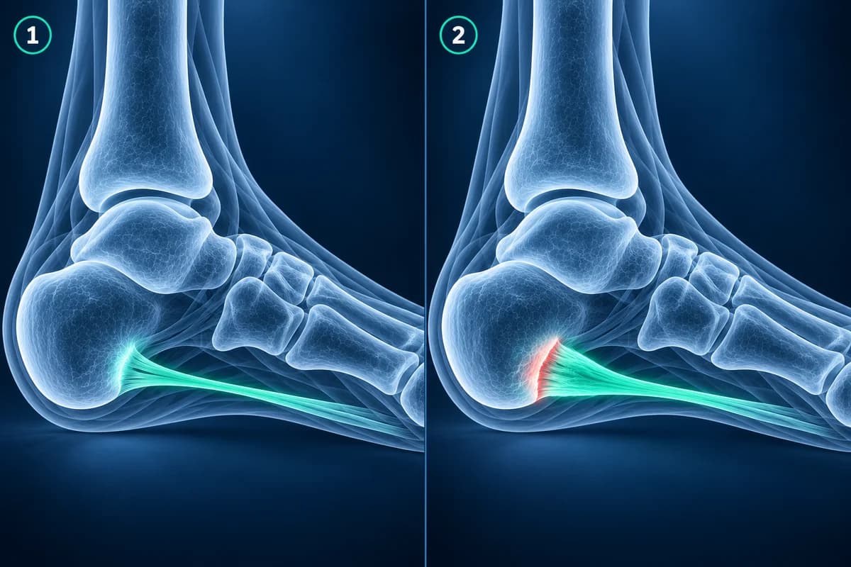
A thick fascia on a follow-up scan does not mean treatment is failing. Thickness falls slowly and often lags behind pain by months, so most clinicians track symptoms and function rather than repeating scans. If you have an MRI study on disc, the free foot MRI viewer opens it privately in your browser, and the guide to reading a foot MRI explains the planes and sequences.
What slows plantar fasciitis recovery?
The common brakes are predictable: a sudden return to running or long walks, long shifts standing on hard floors, worn-out or completely flat shoes, going barefoot on hard surfaces, a very tight calf, and a high body weight. Foot shape plays a part too; both a flat foot and a very high arch change how the fascia is loaded. Pain in both heels together with stiffness in the back or other joints can point to an inflammatory arthritis, and is worth mentioning because it is treated differently.
When is heel pain not routine?
- A sudden pop under the heel with bruising in the sole and difficulty walking
- Heel pain after a fall or jump from height, or pain when squeezing the sides of the heel
- Pain at night or at rest, fever, unexplained weight loss, or a lump in the sole
- Numbness, burning or pins and needles spreading into the sole or toes
- A hot, red, swollen foot in someone with diabetes, even if it does not hurt much
Any of these deserves prompt clinical assessment rather than another month of stretching. A hot, swollen diabetic foot in particular can be an infection or Charcot foot and needs to be seen the same day.
Frequently Asked Questions
Can plantar fasciitis go away on its own?
Often, given enough time, but it tends to be slower and more prone to flares when nothing about the load changes. Simple measures such as calf stretching, better footwear and reducing the activities that provoke pain shorten the course for most people. A clinician or physiotherapist can tailor them to your foot.
Should I stop running with plantar fasciitis?
Not always completely. Many runners reduce distance and pace to a level where heel pain stays mild during the run and is no worse the next morning, then build up gradually. Pain that is clearly worse the morning after a run is a sign the load was too high. Your physiotherapist can help set those limits.
Why is my heel still thick on ultrasound when it no longer hurts?
Tendon and fascia tissue remodels slowly. The thickness of the band near the heel often stays above normal for months after the pain has gone, so the measurement on its own is a poor guide to whether you are recovered. Symptoms and function matter more at that stage.
Does a heel spur need to be removed?
Rarely. A spur on the underside of the heel bone is a reaction to long-term pull at the fascia origin and is found in many people with no heel pain at all. Treatment targets the fascia and its load, and pain usually settles while the spur stays exactly where it is.
When should I see a specialist for heel pain?
When pain has not improved after a few months of consistent treatment, when it is getting worse, when the pattern does not fit, such as pain at rest or numbness, or straight away if any red-flag sign is present. A specialist can confirm the diagnosis, consider imaging and discuss options such as shockwave therapy.
Sources
- Koc TA Jr, Bise CG, Neville C, et al. Heel Pain - Plantar Fasciitis: Revision 2023. J Orthop Sports Phys Ther. 2023;53(12):CPG1-CPG39.
- Buchanan BK, Sina RE, Kushner D. Plantar Fasciitis. StatPearls. Treasure Island (FL): StatPearls Publishing.
- McMillan AM, Landorf KB, Barrett JT, Menz HB, Bird AR. Diagnostic imaging for chronic plantar heel pain: a systematic review and meta-analysis. J Foot Ankle Res. 2009;2:32.
- Expert Panel on Musculoskeletal Imaging. ACR Appropriateness Criteria Chronic Foot Pain: Update 2025. J Am Coll Radiol. 2026;23(4):681-699.
Related Articles
Most plantar fasciitis needs no MRI. Learn when an X-ray, ultrasound or MRI is worth it, what each shows, and what heel pain report phrases mean.
X-ray, MRI, ultrasound or CT for foot pain? What each test shows and misses, which one usually comes first, and how the choice changes for stress fractures, neuromas, plantar fasciitis, Lisfranc injuries and the diabetic foot.
A plain-language guide to your foot MRI: how T1, fluid-sensitive and STIR images differ, which plane shows what, and what bone marrow edema, plantar plate tear and neuroma mean in your report.
Related Conditions
Understand your imaging before your next appointment
MRI, X-ray or CT from a CD, folder or ZIP. A PDF report or photos of the films work too.
Free to open and preview, no account needed. An optional AI report from $10 explains your images in plain language, with questions for your doctor.
Your raw files stay on this device. Only rendered images and minimal context are sent when you request an AI report.
Medical Disclaimer: This page is for informational and educational purposes only. It does not constitute medical advice, diagnosis, or treatment. AI-generated analysis may contain errors. Always consult a qualified healthcare professional for medical decisions. Full Disclaimer