
Ball of Foot Pain Causes: Which Scan Shows What
Ball of foot pain by cause: metatarsalgia, Morton's neuroma, plantar plate tear, stress fracture, Freiberg disease and sesamoiditis, and the test for each.
Key Takeaways
- Metatarsalgia is a location, not a diagnosis; the next step is finding the structure under strain
- Burning between the 3rd and 4th toes fits Morton’s neuroma; ultrasound and MRI are similarly accurate
- Pain under the 2nd toe joint with a drifting toe points to a plantar plate tear
- Stress fractures and early Freiberg disease can hide on the first X-ray but show on MRI
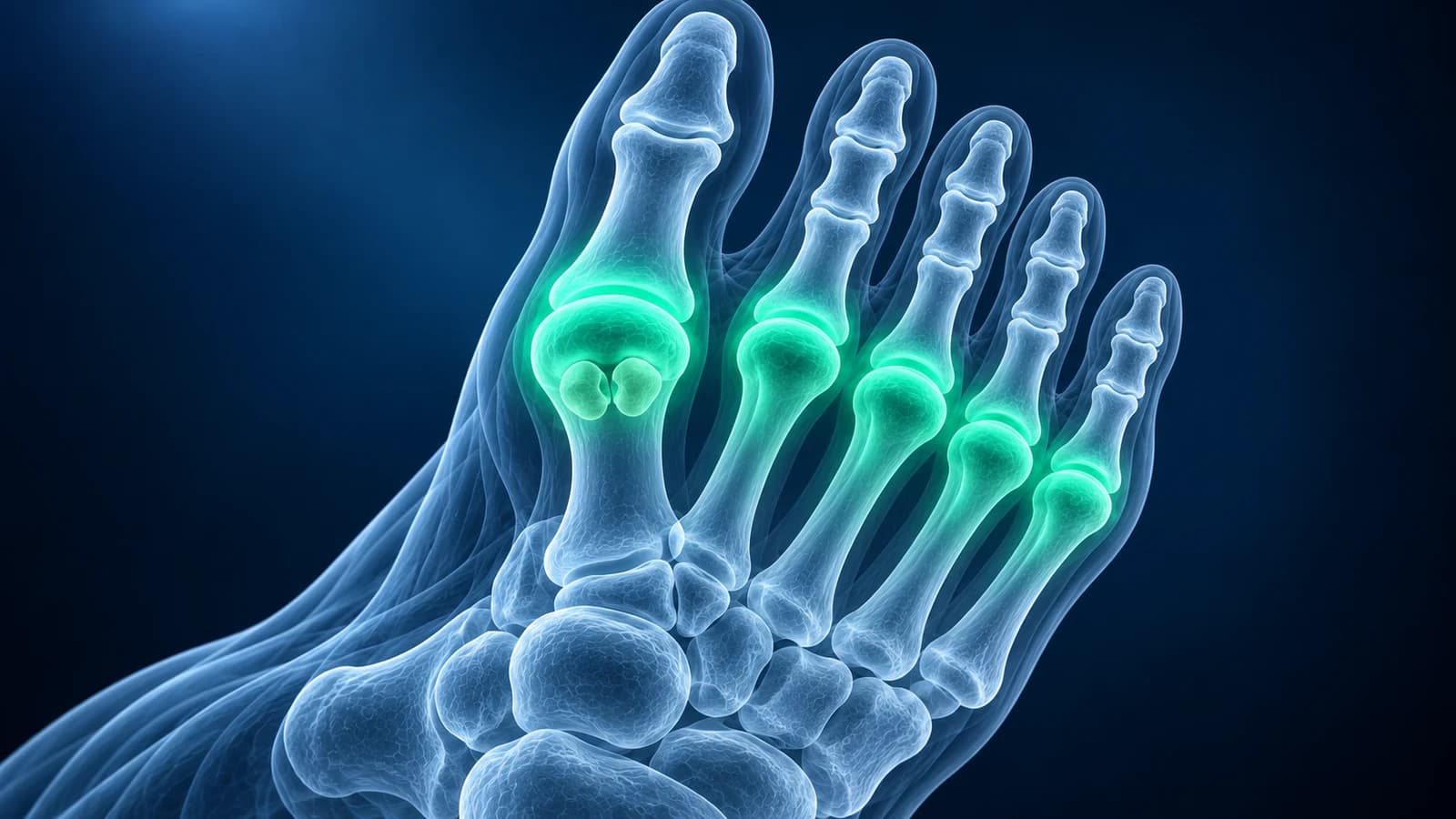
Pain in the ball of the foot is most often overload of the second and third metatarsal heads, called metatarsalgia. Burning between the third and fourth toes suggests Morton’s neuroma, pain under the second toe joint a plantar plate tear, and pain under the big toe the sesamoids. A standing X-ray comes first; ultrasound or MRI follows when it is normal.
“Metatarsalgia” describes where it hurts rather than why, so the real work is narrowing down the structure. This hub shows the usual suspects, the test that sees each one best and the page that covers it in depth. It is educational and does not replace an examination.
Which test shows which cause of ball-of-foot pain?
Following the ACR Appropriateness Criteria for chronic foot pain, foot radiographs are the first test for almost every forefoot complaint. The second test depends on whether the suspect is a bone, a nerve or a ligament.
| Cause | Where it hurts | Test that shows it best | Typical report wording |
|---|---|---|---|
| Metatarsalgia | Under the 2nd and 3rd metatarsal heads, often with callus | Standing X-ray for metatarsal length and alignment | Long second metatarsal, no acute osseous abnormality |
| Morton’s neuroma | Between the 3rd and 4th toes, burning or numb | Ultrasound or MRI, similar accuracy | Intermetatarsal neuroma, intermetatarsal bursitis |
| Plantar plate tear | Under the 2nd toe joint, toe drifting up or sideways | Dynamic ultrasound or MRI | Distal plantar plate tear, MTP joint synovitis |
| Metatarsal stress fracture | On top of and along a metatarsal shaft, swelling | MRI early; X-ray can stay normal for 2 to 4 weeks | Periosteal reaction, marrow edema, fracture line |
| Freiberg disease | Deep in the 2nd toe joint, stiffness | X-ray; MRI in the early stage | Flattening and sclerosis of the 2nd metatarsal head |
| Sesamoiditis | Under the big toe joint, worse pushing off | X-ray with a sesamoid view; MRI if unclear | Sesamoid marrow edema, bipartite or fractured sesamoid |
Which toe is your pain under?
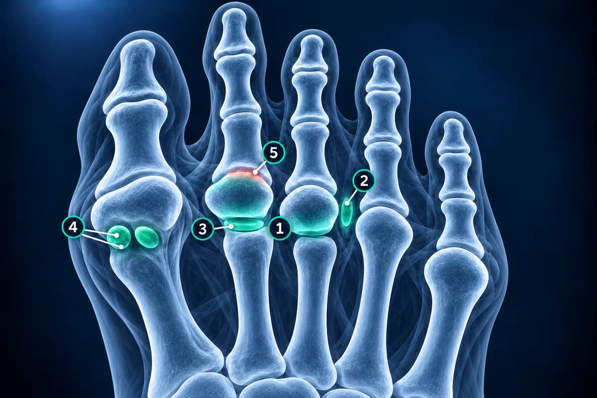
Press along the sole with one finger. Pain directly under a metatarsal head points to the bone or joint. Pain in the soft gap between two heads, especially when you squeeze the forefoot from the sides, points to a nerve. Pain under the big toe that is worse when you bend the toe up points to the sesamoids. Pain on top of the foot along a shaft is more likely a stress fracture.
What is metatarsalgia and how is it imaged?
Metatarsalgia is pressure pain under the middle metatarsal heads, often with a callus over the busiest spot. A second metatarsal that is longer than the first, a bunion that shifts load away from the big toe, high-heeled shoes or a tight calf all push weight onto the middle of the forefoot. A standing X-ray shows the metatarsal lengths and alignment. If it is normal and the pain is focal, ultrasound or MRI looks for the specific structure under strain.
Is it Morton’s neuroma?
A thickened nerve between two metatarsal heads, most often the third and fourth, gives burning, tingling or the feeling of a folded sock under the toes. Ultrasound and MRI detect it with similar accuracy in pooled studies, so many clinicians start with ultrasound; the trade-offs are in Morton’s neuroma: ultrasound vs MRI. Small neuromas also appear on MRI in people without symptoms, so a finding has to match the pain. Details are on the Morton’s neuroma page.
Could the pain under the second toe be a plantar plate tear?
The plantar plate is the thick pad of ligament under each lesser toe joint. When it tears, usually at the second toe, the joint becomes painful and swollen and the toe slowly lifts, crosses over or leaves a gap next to its neighbour. A pooled analysis found dynamic ultrasound more sensitive and MRI more specific for this injury. A plantar plate tear often sits next to a neuroma-like swelling in the same web space, which is why the two are easy to confuse.
Could it be a stress fracture of a metatarsal?
Aching that builds with each walk or run, local swelling on top of the foot and pain when one spot on the shaft is pressed suggest a metatarsal stress fracture, most often in the second or third metatarsal. The first X-ray is frequently normal and can stay normal for 2 to 4 weeks after the pain starts, until new bone forms along the shaft, while MRI shows marrow edema much earlier. Expected healing stages are in the metatarsal fracture healing timeline.
What does a flattened second metatarsal head mean?
A flattened, dense or fragmented head of the second metatarsal on X-ray is the signature of Freiberg disease, a loss of blood supply to that small dome of bone that most often affects teenage girls and young women. In the earliest stage the X-ray can still look normal and MRI shows edema in the head; later stages show collapse and arthritis of the joint, which guides whether treatment is protective or surgical.
Why does it hurt under the big toe joint?
Two sesamoid bones sit inside the tendon under the big toe joint and carry much of the push-off force. Irritation of these bones is sesamoiditis. A special X-ray view looking along the sole shows them side by side. A sesamoid in two pieces may be a fracture or a harmless bipartite sesamoid that formed in two parts; MRI or comparison with the other foot helps tell them apart. Pain on top of the big toe joint with stiffness is a different problem, often hallux rigidus.
When is ball-of-foot pain urgent?
A hot, red, very painful big toe joint that flares overnight can be gout and needs prompt treatment. With diabetes or numb feet, any wound, redness or swelling under the forefoot should be seen the same day because infection can reach the bone without much pain. Read more on gout in the foot.
Can I look at my forefoot images myself?
Yes. MRI series open in our free MRI viewer directly in your browser. Find the short-axis slices, where the metatarsal heads appear as a row of circles, and scroll to the web space or joint named in the report. The sequence names are explained in how to read a foot MRI.
Frequently Asked Questions
What is the most common cause of pain in the ball of the foot?
Overload of the second and third metatarsal heads, called metatarsalgia, is the most common. Morton’s neuroma, plantar plate tears, stress fractures and sesamoid problems are the main specific causes to rule out.
Will an X-ray show Morton’s neuroma?
No. A neuroma is soft tissue and is invisible on X-ray. The X-ray is still useful to exclude a stress fracture, arthritis or Freiberg disease, and ultrasound or MRI is then used to see the nerve itself.
How do I tell a plantar plate tear from Morton’s neuroma?
A plantar plate tear hurts directly under the joint and the toe may drift or lift, while a neuroma hurts between two toes with burning or numbness. The two can coexist, and imaging is often what separates them.
Is ultrasound or MRI better for ball-of-foot pain?
Ultrasound is quick, can test the toe while it moves and is good for neuromas and plantar plates. MRI shows the bone marrow too, so it is better when a stress fracture, Freiberg disease or sesamoid problem is possible.
Can AI explain my forefoot MRI?
AI can match each phrase in a forefoot report to the toe and structure it describes and suggest questions for your clinician. It does not examine your foot and is not a diagnosis.
Sources
- Expert Panel on Musculoskeletal Imaging; Said N, Fox MG, Nacey NC, et al. ACR Appropriateness Criteria Chronic Foot Pain: Update 2025. J Am Coll Radiol. 2026;23(4):681-699.
- Bignotti B, Signori A, Sormani MP, et al. Ultrasound versus magnetic resonance imaging for Morton neuroma: systematic review and meta-analysis. Eur Radiol. 2015;25(8):2254-2262.
- Zanetti M, Strehle JK, Zollinger H, Hodler J. Morton neuroma and fluid in the intermetatarsal bursae on MR images of 70 asymptomatic volunteers. Radiology. 1997;203(2):516-520.
- Albright RH, Brooks BM, Chingre M, et al. Diagnostic accuracy of magnetic resonance imaging (MRI) versus dynamic ultrasound for plantar plate injuries: a systematic review and meta-analysis. Eur J Radiol. 2022;152:110315.
- Carter KR, Chambers AR, Dreyer MA. Freiberg Infraction. StatPearls. Treasure Island (FL): StatPearls Publishing.
- Sims AL, Kurup HV. Painful sesamoid of the great toe. World J Orthop. 2014;5(2):146-150.
- American Academy of Orthopaedic Surgeons. Stress Fractures of the Foot and Ankle. OrthoInfo.
- Koo AY, Tolson DR. March Fracture (Metatarsal Stress Fractures). StatPearls. Treasure Island (FL): StatPearls Publishing.
Related Articles
Which scan finds a Morton's neuroma better, what each one shows and misses, how reports describe it, and when MRI is worth the extra cost.
A plain-language guide to your foot MRI: how T1, fluid-sensitive and STIR images differ, which plane shows what, and what bone marrow edema, plantar plate tear and neuroma mean in your report.
How long a broken metatarsal takes to heal week by week, why the fracture line lingers on X-ray, which fractures heal slowly, and what follow-up reports mean.
Related Conditions
Understand your imaging before your next appointment
MRI, X-ray or CT from a CD, folder or ZIP. A PDF report or photos of the films work too.
Free to open and preview, no account needed. An optional AI report from $10 explains your images in plain language, with questions for your doctor.
Your raw files stay on this device. Only rendered images and minimal context are sent when you request an AI report.
Medical Disclaimer: This page is for informational and educational purposes only. It does not constitute medical advice, diagnosis, or treatment. AI-generated analysis may contain errors. Always consult a qualified healthcare professional for medical decisions. Full Disclaimer