
Metatarsal Fracture Healing Timeline: What Follow-Up X-Rays Show
How long a broken metatarsal takes to heal week by week, why the fracture line lingers on X-ray, which fractures heal slowly, and what follow-up reports mean.
Key Takeaways
- Most middle metatarsal shaft fractures heal in about six to eight weeks
- Callus usually becomes visible on X-ray at two to three weeks
- A faint fracture line on a later film is normal remodeling, not failure to heal
- Jones fractures and proximal fifth metatarsal stress fractures heal slowly and carry a nonunion risk
- MRI finds stress fractures that X-ray misses; CT checks whether a slow fracture has bridged
Most metatarsal fractures heal enough to walk normally in about six to eight weeks, but the X-ray runs behind the foot. Callus usually starts to show at around two to three weeks, a faint fracture line can stay visible for three months or more, and a Jones fracture at the base of the fifth metatarsal often needs twelve weeks or longer.
That gap between how the foot feels and how the film looks is the single most common source of worry after a broken metatarsal. This guide walks through the healing stages week by week, explains the wording follow-up reports use, and lists the fractures that are known to heal slowly. It is general education, not an assessment of your own foot; the clinician treating you decides when to load, when to change footwear and when to repeat imaging.
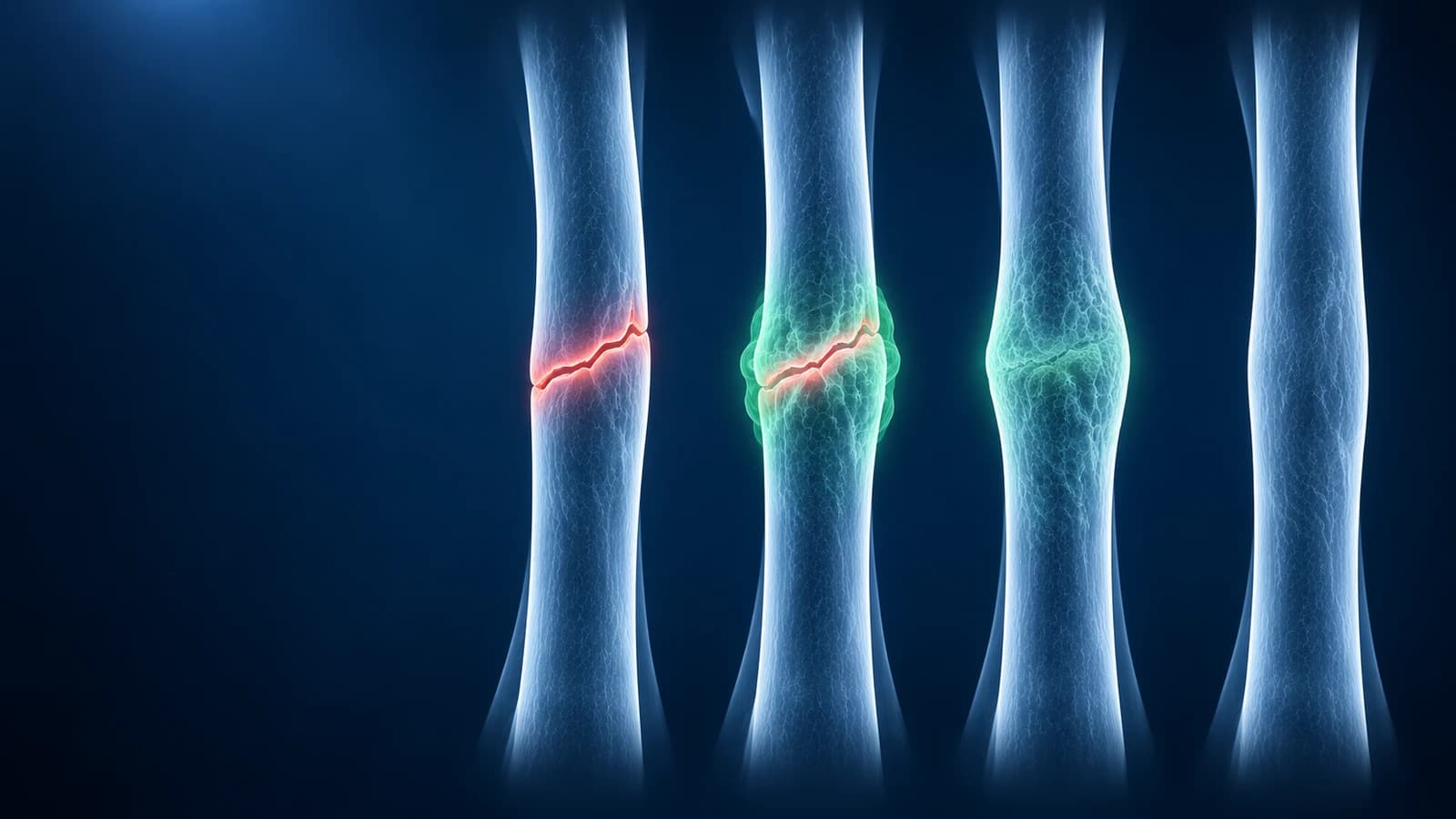
How long does a broken metatarsal take to heal, week by week?
Bone heals in overlapping phases rather than on a fixed calendar, and the forefoot follows the same biology as any long bone. The ranges below describe an uncomplicated, non-displaced shaft fracture of the second, third or fourth metatarsal in a healthy adult. Fractures near the base of the fifth metatarsal, in the first metatarsal, or in people with diabetes or who smoke are slower and are covered further down.
- Days 1 to 7: a blood clot forms between the bone ends and the area swells and bruises, often spreading to the toes and the sole. The X-ray shows only the break itself.
- Weeks 2 to 3: soft callus made of cartilage and woven bone starts to bridge the gap. Pain on walking in a stiff-soled shoe or boot usually eases noticeably, and the edges of the fracture can look slightly blurred on film as the bone ends are resorbed.
- Weeks 4 to 6: the callus mineralises and becomes visible as a cloudy collar around the break. Many people walk in a supportive shoe by now, although the forefoot still aches after a long day.
- Weeks 6 to 12: hard callus bridges the fracture and the bone is described as united, while the line itself may still be faintly visible. Low-impact activity usually returns in this window.
- Months 3 to 12: remodeling trims the callus and restores the normal shaft outline. Swelling at the end of the day and a slightly thicker bone on X-ray are both normal during this phase.
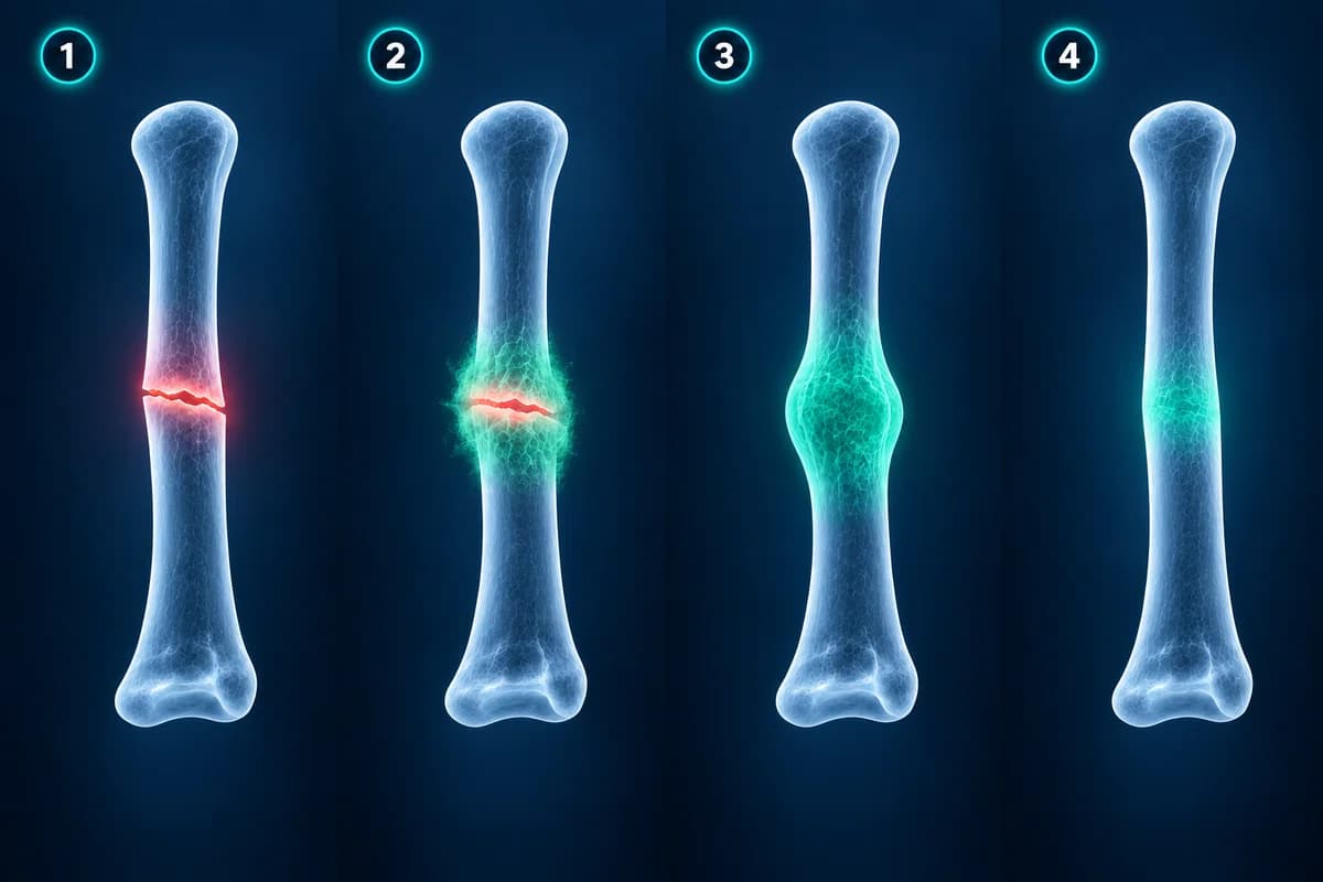
Why does the X-ray still show a fracture line when my foot feels better?
A plain radiograph only sees mineral. Early callus is mostly cartilage and fibrous tissue, which is already stabilising the fracture but is invisible to X-ray until calcium is laid down in it. The opposite happens late in healing: the bone is strong, but the dense line of the original break takes months to fade completely because remodeling is slow. Radiologists therefore talk about healing in terms of callus and bridging rather than whether the line has disappeared.
In practice, clinical union comes first: no tenderness when the bone is pressed and comfortable walking. Radiographic union follows, sometimes weeks later. Most treating clinicians weigh both, and a single film that looks unchanged from the last one is far less meaningful than a trend across two or three visits.
Which metatarsal fractures heal more slowly?
The base of the fifth metatarsal is the classic exception. It is usually divided into three zones. A tuberosity avulsion, where the peroneus brevis tendon pulls off a flake of bone at the very tip, generally heals well with a supportive shoe or boot. A fracture slightly further along, at the junction between the base and the shaft, is the Jones fracture. It sits in a watershed area with a poor blood supply, so it unites slowly and has a meaningful risk of nonunion; treatment is usually a period without weight bearing, or a screw in athletes and in fractures that do not progress. Stress fractures of the proximal shaft behave in a similar way.
Fatigue fractures of the second and third metatarsal shafts are a separate story. These metatarsal stress fractures tend to heal with relative rest and protected walking, but the first X-ray is often normal and the fracture only becomes visible as callus appears two to three weeks later. That delay is explained in our guide to a stress fracture that does not show on X-ray.
Other factors that stretch the timeline include a fracture of the first metatarsal, which carries much of the body's weight at push-off; several metatarsals broken together; displacement or shortening; a break that enters a joint; smoking; diabetes and peripheral neuropathy; low vitamin D or osteoporosis; and returning to impact too early. Multiple fractures at the metatarsal bases also raise the question of a Lisfranc injury of the midfoot, which heals on a very different schedule and is easy to miss on non-weight-bearing films.
What do follow-up X-ray reports mean?
- "Interval callus formation" — new bone has appeared around the break since the previous film, which is the expected sign of healing
- "Periosteal reaction along the shaft" — the bone lining is laying down new bone; in a stress fracture this may be the first visible sign that a fracture was there at all
- "No interval change in alignment" — the fragments have not slipped, angled or shortened since the last visit
- "Bridging callus, fracture line still faintly visible" — the bone is joined across the gap and the remaining line is part of normal remodeling
- "Delayed union" — healing is slower than expected for that fracture and location, but has not stopped
- "Sclerotic fracture margins with persistent lucency" — the bone ends have hardened and a gap remains, a pattern that raises the possibility of nonunion, especially at the fifth metatarsal
If you have the image files from your follow-up visits, you can open the injury film and the latest film side by side in the free foot X-ray viewer in your browser, without uploading anything. The guide to reading a foot X-ray explains the standard views.
When is an MRI or CT scan used during healing?
For an acute foot injury with a suspected occult fracture and normal or unclear X-rays, the ACR rates MRI or CT without contrast as usually appropriate next tests. This addresses a missed initial fracture, not a fixed schedule for checking healing; follow-up imaging is tailored to the fracture and symptoms.
Follow-up of a known metatarsal fracture is done with plain X-rays in almost every case. Further imaging answers a specific question. MRI is used when a stress fracture is suspected but the films are normal: it shows bone marrow edema, the fluid signal inside the bone that appears within days of a stress injury, long before callus. A report might read "bone marrow edema in the third metatarsal shaft with a low-signal fracture line", which means a stress fracture rather than a stress reaction alone.
CT is the tool for the opposite problem: deciding whether a slow fracture has actually bridged. Because it shows the bone in thin slices with no overlap, it can measure how much of the fracture surface has joined, which helps when surgery for a suspected nonunion is being considered. The trade-offs between the three tests are covered in foot MRI vs X-ray vs ultrasound.
When can you walk, drive and run again?
For a stable shaft fracture of the middle metatarsals, many treating teams allow weight bearing as comfort permits in a stiff-soled shoe or walking boot from the start, then a gradual return to normal footwear after four to six weeks. Running and jumping usually wait until the bone is no longer tender, walking is pain free, and the follow-up film shows bridging, which often falls somewhere between eight and twelve weeks. Fifth metatarsal fractures beyond the tuberosity, first metatarsal fractures and fractures treated with surgery follow their own, usually stricter, plans.
Driving depends on which foot is injured, whether you are wearing a boot, and local rules; it is a question for your clinician and, in many countries, your insurer. Pain that returns or increases when you step up activity is the signal to drop back a level and mention it at your next review rather than push through.
When should a healing foot fracture be checked urgently?
- You cannot take four steps on the foot, or pain suddenly worsens after it had been improving
- Toes become pale, blue, cold, numb or tingling
- Severe, bursting pain with tense swelling that is not relieved by elevation
- An open wound over the fracture, or a wound, redness or discharge after surgery
- Calf pain and swelling or sudden breathlessness while you are in a boot or cast
- A warm, red, swollen foot in someone with diabetes or reduced sensation, even when pain is mild
Frequently Asked Questions
Can a broken metatarsal heal without a cast?
Many can. Non-displaced shaft fractures of the second to fourth metatarsals, and avulsion fractures at the tip of the fifth metatarsal tuberosity, are commonly treated with a stiff-soled shoe or a walking boot and weight bearing as comfort allows. Fractures of the first metatarsal, displaced fractures, several broken metatarsals and Jones fractures are more often protected more strictly. Which group a fracture belongs to is a decision for the clinician who has examined the foot and reviewed the films.
Is it normal for my foot to swell months after a metatarsal fracture?
Yes. The forefoot sits at the lowest point of the body, so swelling at the end of the day often continues for three to six months after the bone has united, and the healed metatarsal can stay slightly thicker on X-ray for a year. Swelling that is increasing, hot, red, or paired with new pain is different and should be reviewed.
What is the difference between delayed union and nonunion?
Delayed union means the fracture is healing more slowly than expected for its type and location but is still progressing. Nonunion means healing has stalled, typically shown by a persistent gap with hardened, rounded bone edges on serial X-rays or CT. Nonunion is uncommon in the middle metatarsals and is mainly a concern at the proximal fifth metatarsal, where it is sometimes treated with surgery.
How many follow-up X-rays are usual?
It varies. A stable fracture may have one check film or none, while a fracture that could shift is often re-imaged at one to two weeks to confirm the position held, and a slow-healing fracture is followed every few weeks until it bridges. Fewer films is not a sign of less care; it usually means the fracture is predictable.
Does the AI tool decide when I can walk on my foot?
No. An AI explanation can describe what is visible on your images and translate the report wording into plain language, which may help you prepare questions. It is not a diagnosis and cannot examine the foot, so decisions about loading, footwear and return to sport stay with your treating clinician.
Sources
- Hatch RL, Alsobrook JA, Clugston JR. Diagnosis and management of metatarsal fractures. Am Fam Physician. 2007;76(6):817-826.
- Bowes J, Buckley R. Fifth metatarsal fractures and current treatment. World J Orthop. 2016;7(12):793-800.
- Bergman R, Kaiser K. Stress Reaction and Fractures. StatPearls. Treasure Island (FL): StatPearls Publishing.
Related Articles
Why a foot stress fracture can hide on a normal X-ray, what MRI, CT and repeat X-rays show instead, and which foot stress fractures are high risk.
A plain-language guide to your foot X-ray: the three standard views, how to trace alignment from heel to toes, and what fractures, fleck signs, accessory bones and arthritis look like.
X-ray, MRI, ultrasound or CT for foot pain? What each test shows and misses, which one usually comes first, and how the choice changes for stress fractures, neuromas, plantar fasciitis, Lisfranc injuries and the diabetic foot.
Related Conditions
Understand your imaging before your next appointment
MRI, X-ray or CT from a CD, folder or ZIP. A PDF report or photos of the films work too.
Free to open and preview, no account needed. An optional AI report from $10 explains your images in plain language, with questions for your doctor.
Your raw files stay on this device. Only rendered images and minimal context are sent when you request an AI report.
Medical Disclaimer: This page is for informational and educational purposes only. It does not constitute medical advice, diagnosis, or treatment. AI-generated analysis may contain errors. Always consult a qualified healthcare professional for medical decisions. Full Disclaimer