
Freiberg Disease (Infraction): Second Metatarsal Head Collapse on Imaging
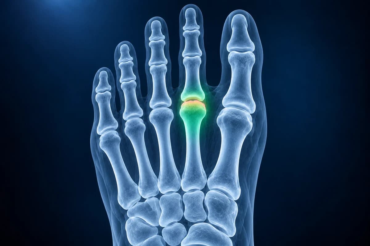
Freiberg disease, also called Freiberg infraction, is a collapse of the rounded head of a metatarsal, almost always the second, after part of its bone loses blood supply. Early X-rays can be normal while MRI already shows marrow change; later films show the head flattening, sclerosis and eventually arthritis of the joint at the base of the toe.
Common Symptoms
- Pain under the ball of the foot at the base of the second toe, less often the third
- Pain that is worse when walking barefoot, running, jumping or wearing high heels
- Swelling and tenderness around the joint where the toe meets the foot
- Stiffness and reduced bending of the affected toe, especially upward
- A feeling of walking on a pebble or a bony lump under the forefoot in later stages
- Symptoms that often begin in the teenage years and are more common in girls and women
What causes Freiberg disease?
Freiberg disease belongs to a group of conditions called osteochondroses, in which a growing or recently grown area of bone and its cartilage are damaged by reduced blood flow. The exact cause is not settled. The leading explanation is repeated stress on the metatarsal head while the bone is still maturing, which leads to tiny fractures beneath the joint surface, interrupted blood supply, and then death and collapse of part of the bone. It was first described by Albert Freiberg in 1914, and the older word "infraction" refers to that small fracture.
The second metatarsal is a natural target. It is often the longest metatarsal and is locked firmly at its base, so it takes high pressure at toe-off. A first metatarsal that is short or bends upward easily, a bunion that shifts load away from the big toe, and frequent high heels or barefoot sports all increase that pressure. It is most often diagnosed in teenage girls and young women, though adults can present with the late, arthritic stage.
What does Freiberg disease look like on X-ray?
For chronic forefoot pain, the ACR Appropriateness Criteria place standing radiographs first. In the earliest stage the X-ray may be normal or show only slight widening of the joint space from fluid. Over the following weeks to months, the top of the metatarsal head becomes denser (sclerosis), then a dent or flattening appears in its rounded surface, sometimes with a crescent of lucency just under the cartilage. In longer-standing disease the head looks broad and flat, the bone behind it thickens, loose fragments may sit in the joint, and bony spurs form, which is the picture of secondary arthritis.
Comparing the head with its neighbours on the same film is often the easiest way to see the change. You can do this on your own images in the free foot X-ray viewer, and our guide on how to read a foot X-ray explains what each view shows.
When is MRI used for Freiberg disease?
MRI is most useful when pain is localised to a metatarsal head but X-rays are normal or unclear. It shows bone marrow edema in the head, low signal beneath the joint surface on T1-weighted images where bone has died, and fluid or thickened lining in the joint. MRI also helps separate Freiberg disease from conditions that cause pain in the same spot. Our article on foot MRI vs X-ray vs ultrasound explains when each test adds information, and you can scroll a scan in the free foot MRI viewer.
What do Freiberg report phrases mean?
- "Flattening of the second metatarsal head"
- The normally dome-shaped end of the bone has partly collapsed. This is the hallmark X-ray sign.
- "Subchondral sclerosis" or "subchondral lucency"
- Denser or thinner-looking bone just beneath the cartilage, reflecting dead bone and the body's attempt to repair it.
- "Osteonecrosis" or "avascular necrosis of the metatarsal head"
- Medical terms for bone that has died from lost blood supply, the process behind Freiberg disease.
- "Intra-articular loose body"
- A fragment of bone or cartilage that has broken off and sits inside the joint, which can cause catching.
- "Secondary degenerative change of the second MTP joint"
- Arthritis that has developed because the joint surface is no longer smooth, typically seen in late stages.
What are the stages of Freiberg disease?
The Smillie classification, still quoted in reports and surgical reviews, describes five stages. Stage 1 is a fine crack in the head that may show only on MRI. Stage 2 is early collapse of the central joint surface. Stage 3 is deeper central collapse with the edges of the head still holding. Stage 4 adds a loose fragment in the joint. Stage 5 is end-stage arthritis with a flattened, deformed head. Earlier stages have the best chance of settling with offloading, while later stages more often need surgery. Use the X-ray report phrase guide to place Freiberg staging terms alongside other findings.
What we look for on metatarsal head imaging
- Shape of the second and third metatarsal heads compared with the neighbouring heads
- Sclerosis, subchondral lucency or a crescent line beneath the joint surface
- Joint space width, loose fragments and spur formation at the MTP joint
- Marrow edema and T1 low signal in the metatarsal head on MRI
- Relative metatarsal lengths and first ray position that increase second-toe loading
What else causes pain under the second toe?
Pain under the second metatarsal head is common and has several causes. A plantar plate tear causes pain and swelling with the toe drifting or lifting. A Morton's neuroma gives burning or numbness between the toes. A metatarsal stress fracture tends to hurt along the shaft rather than at the joint. Inflammatory arthritis and gout can inflame the same joint. Imaging, the examination and sometimes blood tests separate them.
Seek prompt care if the joint becomes hot, red and very swollen, especially with fever, or if you have diabetes and notice a wound, colour change or new swelling in the forefoot. Those signs suggest infection or another problem that should not wait for a routine appointment.
How is Freiberg disease treated?
Treatment starts with taking pressure off the metatarsal head: a stiff-soled or rocker-bottom shoe, a metatarsal pad placed just behind the painful head, avoiding high heels and barefoot impact, and sometimes a walking boot or short period of reduced weight bearing during a flare. Pain relief and activity changes are added as needed. Many people, particularly teenagers caught early, settle with this approach.
When pain persists or the head has collapsed, surgery may be offered. Options include cleaning out the joint and removing loose bodies, a wedge osteotomy that rotates healthier cartilage into the weight-bearing area, shortening the metatarsal to reduce load, and, for advanced arthritis, procedures that reshape or replace part of the joint. A foot and ankle surgeon chooses based on the stage and your symptoms.
Can AI explain my forefoot X-ray or MRI?
Upload your images and report for a plain-language AI explanation of the metatarsal head shape, the report terms and what to ask at your appointment. It is an educational review, not a diagnosis, and does not replace the clinician who examines your foot. Use the ball-of-foot pain guide when discussing where Freiberg symptoms occur.
When should forefoot pain be checked urgently?
Seek urgent medical advice if you cannot walk, your foot changes shape, or it becomes hot, swollen and painful, especially if you feel feverish or shivery. Do not assume these symptoms are simply Freiberg disease.
Frequently Asked Questions
Is Freiberg disease the same as Freiberg infraction?
Yes. Both names describe the same condition: collapse of a metatarsal head, usually the second, after part of the bone loses its blood supply. "Infraction" is the older term and reflects the idea that small fractures start the process.
Can Freiberg disease heal on its own?
In early stages, especially in adolescents, the bone can revascularise and symptoms often settle with offloading over months. Once the head has flattened, its shape does not return to normal, but many people still become comfortable with footwear changes. Persistent pain in later stages is the usual reason for surgery.
Why was my X-ray normal if I have Freiberg disease?
The earliest stage is a change in the bone marrow and a tiny crack that X-rays cannot see. Flattening and sclerosis appear only after the bone has started to collapse. MRI or a repeat X-ray after some weeks is often used when symptoms point to the second metatarsal head.
Can I keep running with Freiberg disease?
High-impact running and jumping usually aggravate it, so most plans include a break from them during painful phases and a gradual return in stiff or rocker-soled shoes. Low-impact activities such as cycling or swimming are often tolerated. Your clinician can advise based on the stage seen on your imaging.
Sources
- Carter KR, Chambers AR, Dreyer MA. Freiberg Infraction. StatPearls. Treasure Island (FL): StatPearls Publishing.
- Cerrato RA. Freiberg's disease. Foot Ankle Clin. 2011;16(4):647-658.
- Expert Panel on Musculoskeletal Imaging; Said N, Fox MG, Nacey NC, et al. ACR Appropriateness Criteria Chronic Foot Pain: Update 2025. J Am Coll Radiol. 2026;23(4):681-699.
- NHS. Pain in the ball of the foot.
Related Articles
Why a foot stress fracture can hide on a normal X-ray, what MRI, CT and repeat X-rays show instead, and which foot stress fractures are high risk.
Which scan finds a Morton's neuroma better, what each one shows and misses, how reports describe it, and when MRI is worth the extra cost.
Understand your imaging before your next appointment
MRI, X-ray or CT from a CD, folder or ZIP. A PDF report or photos of the films work too.
Free to open and preview, no account needed. An optional AI report from $10 explains your images in plain language, with questions for your doctor.
Your raw files stay on this device. Only rendered images and minimal context are sent when you request an AI report.
Medical Disclaimer: This page is for informational and educational purposes only. It does not constitute medical advice, diagnosis, or treatment. AI-generated analysis may contain errors. Always consult a qualified healthcare professional for medical decisions. Full Disclaimer