
Foot X-Ray Report Phrases Explained in Plain Language
What hallux valgus angle, fleck sign, joint space narrowing at the first MTP, pes planus, calcaneal spur and Jones fracture mean on your foot X-ray report.
Key Takeaways
- A hallux valgus angle below 15° and an intermetatarsal angle below 9° are counted as normal
- A fleck sign is a small chip that marks a Lisfranc ligament injury and needs follow-up
- Joint space narrowing with a dorsal spur at the big toe joint fits hallux rigidus
- A heel spur is common without pain and is rarely the treatment target
- Zone 1 fifth metatarsal fractures usually heal readily; zone 2 Jones fractures heal more slowly
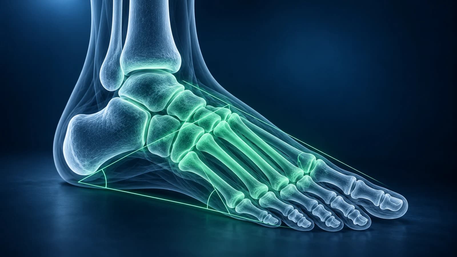
Foot X-ray reports use a small set of phrases: angles that describe alignment, words for small bone fragments, and terms for wear in a joint. “Hallux valgus angle 28°, intermetatarsal angle 13°”, for instance, means a moderate bunion, because a hallux valgus angle below 15° and an intermetatarsal angle below 9° are counted as normal. Each phrase below is explained with what to ask next.
X-ray shows bone and alignment well but does not show ligaments, nerves, cartilage or bone swelling directly, so a “normal” report does not rule everything out. This page explains wording only and is not a diagnosis of your foot.
Where on a foot X-ray do the common report phrases point?
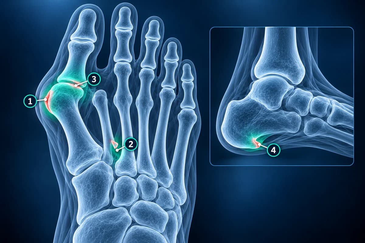
A standard foot series has three pictures: top-down (dorsoplantar), angled (oblique) and side (lateral). Angles for bunions and flat feet are only reliable on films taken standing, so look for “weight-bearing” in the report header. If it says “non-weight-bearing”, the alignment numbers may understate the problem.
What does “hallux valgus angle 30°, intermetatarsal angle 14°” mean?
The hallux valgus angle (HVA) is how far the big toe leans toward the second toe; the intermetatarsal angle (IMA) is how far the first metatarsal has drifted away from the second. Together they grade a bunion: in a widely used scheme an HVA of 20° to 40° with an IMA of 11° to 16° is moderate, and values above those are severe. So this example describes a moderate deformity. Reports may also mention “lateral sesamoid subluxation”, meaning the sesamoids have slid out from under the metatarsal head.
Ask your clinician: were these films taken standing, and does the size of the angle change which treatment you would suggest? How each line is drawn is shown step by step in foot X-ray angles explained.
What does “fleck sign” mean on a foot X-ray?
It is a tiny chip of bone lying in the gap between the base of the first and second metatarsals. The chip has been pulled off by the Lisfranc ligament, so although it looks trivial it is a marker of a Lisfranc injury. Reports sometimes add “widening of the first intermetatarsal space” or “malalignment of the second tarsometatarsal joint”, which strengthen the suspicion.
Ask your clinician: should I avoid putting weight on the foot until this is clarified, and do I need standing films of both feet, a CT or an MRI? Why these injuries are often missed on first films is covered in Lisfranc injury missed on X-ray.
What does “joint space narrowing at the first MTP joint” mean?
The first MTP joint is the big toe joint. Cartilage does not show on X-ray, so the gap between the bones stands in for it; a narrower gap means thinner cartilage. When the report adds “dorsal osteophyte” (a spur on top of the joint) and “subchondral sclerosis” (whiter bone under the surface), the pattern fits hallux rigidus, arthritis that stiffens the toe. Sharp-edged “punched-out erosions” beside the same joint point instead toward gout.
Ask your clinician: is this wear or inflammation, how much movement is left in the joint, and would a stiff-soled shoe or rocker sole help before anything else?
What do “pes planus” and “Meary’s angle” mean?
Pes planus is the medical name for flat foot. Meary’s angle is drawn on the standing side view along the talus and the first metatarsal; in a typical arch the two lines run almost straight on. When the line breaks downward toward the sole, the arch has dropped; when it breaks upward, the report may say “pes cavus”, a high arch. A rigid flat foot in a teenager can come from a bony or fibrous bar between two tarsal bones, called tarsal coalition.
Ask your clinician: is the flat foot flexible or rigid, does it hurt, and is the tendon on the inner ankle involved? A painless flexible flat foot often needs no treatment.
Is a “calcaneal spur” the cause of heel pain?
Often not on its own. A plantar calcaneal spur is a small bony beak under the front of the heel bone, seen on the side view. Pooled studies find spurs more often in people with heel pain, but many people without any pain have them too, and removing a spur is rarely the treatment. The pain usually comes from the band of tissue that attaches next to it, in plantar fasciitis. A spur at the back of the heel, where the Achilles tendon attaches, is a different finding.
Ask your clinician: does the spur change anything about treatment, or is the plan the same as for plantar fasciitis without one?
What is the difference between a Jones fracture and a pseudo-Jones fracture?
Both are breaks at the base of the fifth metatarsal, on the outer edge of the foot, and the report names the zone. Zone 1 is a chip pulled off the very tip (the tuberosity), often called a “pseudo-Jones” or avulsion fracture; it usually heals well in a supportive shoe or boot. Zone 2, a little further along where the blood supply is poorer, is the true Jones fracture, which heals more slowly. Zone 3, in the shaft just beyond, is usually a stress fracture.
Ask your clinician: which zone is it, how long should I stay off the foot, and is surgery discussed because of my sport or activity level? The expected timeline is set out in metatarsal fracture healing timeline.
What happens after the X-ray if the report is normal or unclear?
The American College of Radiology places X-ray first for most foot injuries and for chronic foot pain. After an injury, the Ottawa foot rules help decide whether an X-ray is needed at all: pain in the midfoot plus tenderness at the base of the fifth metatarsal or the navicular, or being unable to take four steps. If the films are normal but pain persists, MRI is usually next for a suspected stress fracture or soft-tissue injury, CT for complex midfoot or heel fractures, and ultrasound for a neuroma or plantar fascia. A repeat X-ray a few weeks later can also reveal a stress fracture that was invisible at first.
Which foot X-ray findings need prompt care?
Phrases such as “displaced fracture”, “dislocation”, “gas in the soft tissues”, “cortical destruction” or “fragmentation” of midfoot bones in someone with diabetes need prompt medical contact, especially with fever, a wound, or a foot that is hot, red and swollen. These can signal infection or Charcot foot, where early treatment matters.
Can I measure the angles on my own X-ray?
Yes, roughly. Open the DICOM files in our free X-ray viewer, which works in your browser without uploading the files, and use the angle tool along the shafts of the big toe and first metatarsal. Expect a few degrees of difference from the report, which is normal between readers. For the views and landmarks, see how to read a foot X-ray.
Frequently Asked Questions
What does “no acute osseous abnormality” mean?
It means no fresh fracture or dislocation was seen. It does not rule out a ligament sprain, an early stress fracture or a soft-tissue problem, which X-ray cannot show directly.
What is an “os peroneum” or “accessory ossicle”?
It is a small extra bone that many people are born with. It is usually harmless, but it can be mistaken for a fracture fragment, and an extra bone beside the navicular can become painful as an accessory navicular.
What does “periosteal reaction” on a metatarsal mean?
It is new bone forming along the outside of the shaft, most often because a stress fracture is healing. Less often it comes from infection or other causes, which your clinician weighs against your symptoms.
Why do two reports give different angles for my foot?
Angles depend on where the lines are drawn and on whether the foot was standing. A difference of a few degrees between readers or between visits is common and usually does not change the grade.
Can AI read my foot X-ray report?
AI can explain the terms and angles in plain language and help you prepare questions. It does not replace the radiologist or the clinician who examines your foot and decides on treatment.
Sources
- Expert Panel on Musculoskeletal Imaging; Gorbachova T, Chang EY, Ha AS, et al. ACR Appropriateness Criteria Acute Trauma to the Foot. J Am Coll Radiol. 2020;17(5S):S2-S11.
- Expert Panel on Musculoskeletal Imaging; Said N, Fox MG, Nacey NC, et al. ACR Appropriateness Criteria Chronic Foot Pain: Update 2025. J Am Coll Radiol. 2026;23(4):681-699.
- Ray JJ, Friedmann AJ, Hanselman AE, et al. Hallux Valgus. Foot Ankle Orthop. 2019;4(2):2473011419838500.
- Kalia V, Fishman EK, Carrino JA, et al. Epidemiology, imaging, and treatment of Lisfranc fracture-dislocations revisited. Skeletal Radiol. 2012;41(2):129-136.
- Lawrence SJ, Botte MJ. Jones' fractures and related fractures of the proximal fifth metatarsal. Foot Ankle. 1993;14(6):358-365.
- McMillan AM, Landorf KB, Barrett JT, Menz HB, Bird AR. Diagnostic imaging for chronic plantar heel pain: a systematic review and meta-analysis. J Foot Ankle Res. 2009;2:32.
- Younger AS, Sawatzky B, Dryden P. Radiographic assessment of adult flatfoot. Foot Ankle Int. 2005;26(10):820-825.
- Bachmann LM, Kolb E, Koller MT, et al. Accuracy of Ottawa ankle rules to exclude fractures of the ankle and mid-foot: systematic review. BMJ. 2003;326(7386):417.
Related Articles
A plain-language guide to your foot X-ray: the three standard views, how to trace alignment from heel to toes, and what fractures, fleck signs, accessory bones and arthritis look like.
What the numbers on your foot X-ray mean: hallux valgus angle, intermetatarsal angle, Meary's angle, calcaneal pitch and Böhler's angle, with normal ranges and how each is measured.
Why Lisfranc midfoot injuries are missed on first X-rays, the alignment lines and fleck sign to check, and when standing X-rays, CT or MRI are needed.
Related Conditions
Understand your imaging before your next appointment
MRI, X-ray or CT from a CD, folder or ZIP. A PDF report or photos of the films work too.
Free to open and preview, no account needed. An optional AI report from $10 explains your images in plain language, with questions for your doctor.
Your raw files stay on this device. Only rendered images and minimal context are sent when you request an AI report.
Medical Disclaimer: This page is for informational and educational purposes only. It does not constitute medical advice, diagnosis, or treatment. AI-generated analysis may contain errors. Always consult a qualified healthcare professional for medical decisions. Full Disclaimer