
Metatarsalgia: Finding the Cause of Ball-of-Foot Pain on Imaging
Metatarsalgia is a description, not a diagnosis: it means pain under the ball of the foot, usually beneath the second, third and fourth metatarsal heads. Imaging is used to find what is behind it. A standing X-ray comes first; MRI or ultrasound is added when the X-ray is normal and the pain persists or points to one joint.
Common Symptoms
- Aching or burning pain under the ball of the foot, usually beneath the second, third or fourth toe
- A feeling of walking on a pebble or a folded sock under the forefoot
- Pain that grows when standing, walking barefoot on hard floors, running or wearing high heels
- Thick callus under one or more metatarsal heads where the skin takes the most pressure
- Relief when sitting down or switching to a cushioned, stiff-soled shoe
- Tenderness when the area under a single toe joint is pressed with a fingertip
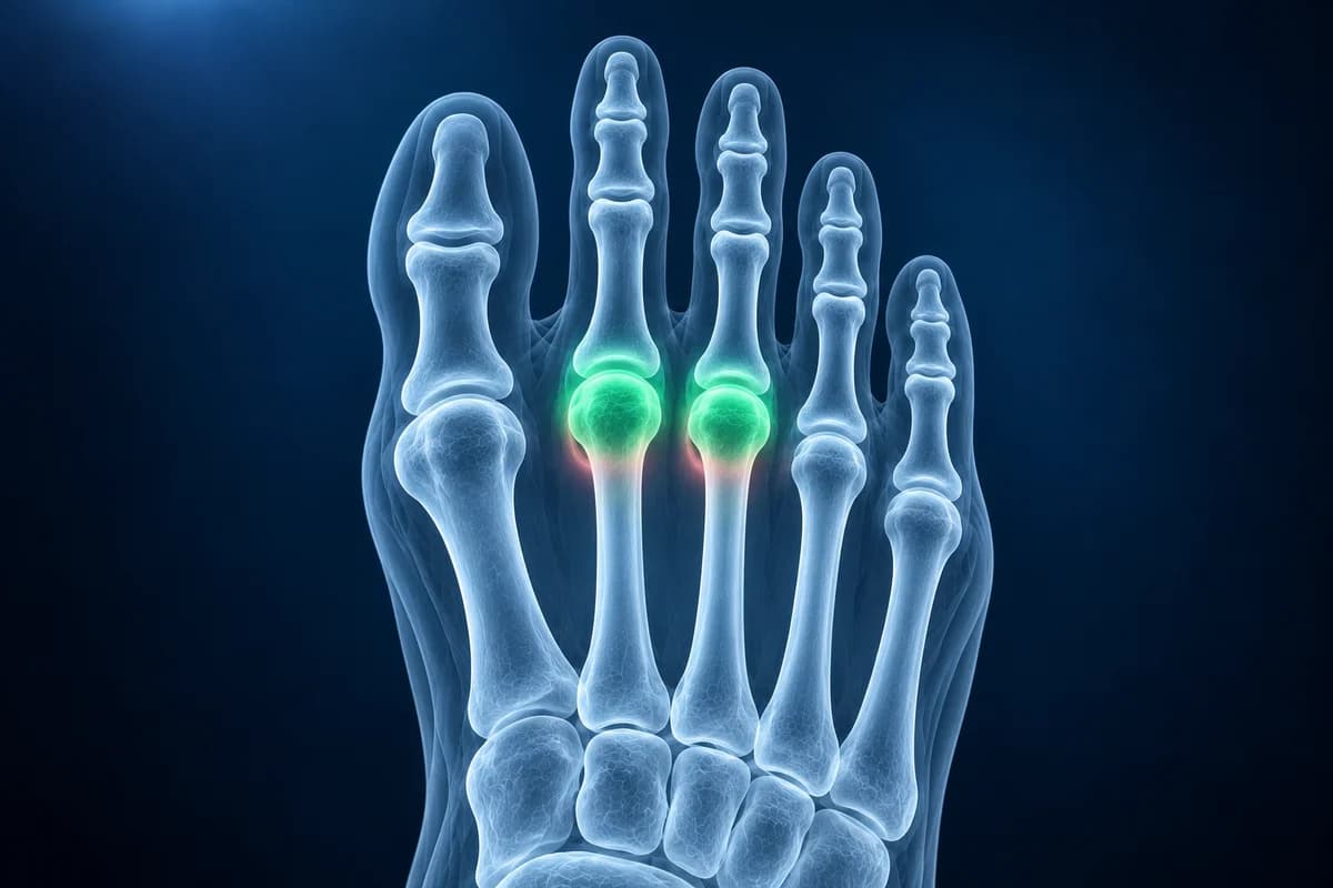
The ball of the foot is where the long metatarsal bones meet the toes. Every step rolls the body over these joints, so overload, a bone stress injury, a torn ligament, a pinched nerve or an inflamed joint lining can all produce the same complaint. This page explains which of those causes each test can find, the wording that turns up on forefoot reports, and when ball-of-foot pain should be seen quickly. It is educational information, not a diagnosis.
What causes pain under the ball of the foot?
Reviews of metatarsalgia, such as the one by Besse, separate primary causes, where the foot shape itself overloads a metatarsal head, from secondary causes, where another condition does it, and from iatrogenic causes left by earlier foot surgery. The imaging work-up is really a search through this list:
- Mechanical overload: a second metatarsal that is long compared with the first, a high arch, a tight calf, a bunion that shifts weight sideways, or a thinning fat pad under the forefoot with age.
- Bone stress: a metatarsal stress fracture or the earlier stress reaction, typically after a jump in walking or running.
- A flattened metatarsal head from Freiberg disease, where part of the second or third head loses its blood supply and collapses.
- Instability of a toe joint from a plantar plate tear, the ligament under the joint that stops the toe from drifting upward.
- Nerve pain between two metatarsal heads from a Morton's neuroma, felt as burning or numbness spreading into two neighbouring toes.
- Inflammation of the toe joint lining (MTP synovitis), either on its own from overload or as an early sign of gout or rheumatoid arthritis.
Pain under the big toe joint is usually discussed separately, because the sesamoid bones there have their own set of problems. A broader walk-through of forefoot causes and the tests for each is in the guide to ball-of-foot pain causes and imaging.
Do you need an X-ray for metatarsalgia?
If the pain has lasted more than a few weeks, an X-ray is the usual first test. The American College of Radiology Appropriateness Criteria for chronic foot pain start with radiographs, and foot surgery reviews of metatarsalgia advise taking them standing, because toe alignment and metatarsal length change under body weight. The X-ray answers several questions at once: is there a healing stress fracture, a collapsed metatarsal head, joint narrowing from arthritis, a toe base that has slid upward on its metatarsal, or a bunion crowding the lesser toes?
What an X-ray cannot show is soft tissue: the plantar plate, the nerve, the fat pad and inflamed joint lining are invisible on it, and an early stress reaction often looks normal for the first weeks. A normal X-ray therefore does not mean nothing is wrong. You can review your own films in the free foot X-ray viewer and compare the length of the first and second metatarsals.
When is an MRI or ultrasound added for ball-of-foot pain?
When the X-ray is normal or unclear and the pain does not settle with shoe changes and offloading, the ACR criteria list MRI and ultrasound as the next steps, chosen by the question being asked.
- MRI sees bone and soft tissue together. It is the better choice when a stress reaction, an early Freiberg collapse or several possible causes are in play, because bone marrow edema shows on MRI long before an X-ray changes.
- Ultrasound is quick and lets the examiner press on the painful spot and move the toe while watching. It suits a focused question such as a suspected neuroma, a plantar plate tear or joint fluid, but it shows little of the bone marrow.
- CT is rarely needed for metatarsalgia. It is mainly used to map bone before surgery that changes metatarsal length.
If you already have an MRI on disc or as DICOM files, the free foot MRI viewer lets you scroll through the forefoot slices; the guide to reading a foot MRI explains which planes show the metatarsal heads best.
What does “bone marrow edema in the second metatarsal head” mean on your MRI report?
Forefoot reports rarely use the word metatarsalgia. They describe what was found, and each phrase below points toward a different cause. Use the question after each one to guide the conversation with your clinician.
- “Bone marrow edema in the second metatarsal”: extra fluid inside the bone, a sign of stress or overload that can come before a fracture line appears. Ask whether it fits a stress reaction and how long to reduce impact.
- “Flattening of the metatarsal head with subchondral sclerosis”: the dome of the head has partly collapsed, the pattern of Freiberg disease. Ask what stage it is and whether the joint surface is still smooth.
- “Plantar plate attenuation at the second MTP joint”: the ligament under the toe joint is thinned or starting to tear. Ask whether the toe is unstable on examination and whether taping or a stiff shoe is enough.
- “Intermetatarsal space lesion” or “intermetatarsal bursitis”: a small mass or fluid pocket between two metatarsal heads, which may be a neuroma. Ask whether it matches where your symptoms are, because these are also seen in people without pain.
- “MTP joint effusion and synovitis”: fluid and thickened lining in a toe joint. Ask whether it looks like overload alone or whether blood tests for gout or inflammatory arthritis are worthwhile.
- “Relatively long second metatarsal”: an X-ray finding where the second metatarsal reaches further forward than the first, putting more load on its head. Ask whether insoles or shoe changes are expected to help before anything else is considered.
More forefoot wording, from “stress reaction” to “sesamoid edema”, is translated in foot MRI report phrases explained.
What We Look For Under the Ball of the Foot
- Relative length of the first, second and third metatarsals on the standing top-down view
- Bone marrow edema, a fracture line or new bone along a metatarsal shaft or neck
- Shape of each metatarsal head, including flattening, fragmentation or joint narrowing
- Position of each toe base on its metatarsal head and signs of upward subluxation
- Plantar plate continuity, joint fluid and synovitis at the lesser toe joints
- Masses or fluid between the metatarsal heads and the thickness of the forefoot fat pad
How is metatarsalgia treated once the cause is known?
Most ball-of-foot pain is treated without surgery. The usual first steps are a shoe with a wide toe box, a stiff or rocker sole and a lower heel; a metatarsal pad placed just behind the painful heads rather than under them; calf stretching when the ankle is tight; and a temporary cut in impact. Callus over a pressure point is trimmed so it stops acting like a stone in the shoe.
The imaging result changes what comes next. A stress fracture needs a period of protected walking, Freiberg disease or a torn plantar plate may need a boot or later surgery, and synovitis from gout or rheumatoid arthritis needs treatment of the underlying disease. When a long metatarsal keeps overloading despite good shoes and insoles, surgeons sometimes shorten it with an osteotomy, a decision that is made on the standing X-ray and the examination together.
When is ball-of-foot pain a reason to be seen quickly?
- You have diabetes or reduced feeling in the feet and notice a blister, a crack, bleeding into a callus or a sore under a metatarsal head. Pressure points like these can turn into ulcers and infection.
- The forefoot becomes hot, red and swollen, or you have a fever.
- You cannot put weight on the foot, or there is swelling and bruising on top of the forefoot after a run.
- Several toe joints in both feet are swollen and stiff in the morning, which can point to inflammatory arthritis.
AI can help explain the forefoot findings described in your report, but it cannot press on the painful spot, test the toe joints or look at your shoes. Treat its output as preparation for a visit, not as a verdict.
Frequently Asked Questions
Is metatarsalgia a diagnosis?
It is a name for a symptom: pain under the metatarsal heads. A clinician aims to find the cause behind it, such as overload, a stress fracture, Freiberg disease, a plantar plate tear or a neuroma, because each one is treated differently.
Can metatarsalgia show up on an X-ray?
The pain itself does not, but many of its causes do: a long second metatarsal, a flattened metatarsal head, a healing stress fracture, arthritis or a toe that has slid out of joint. A normal standing X-ray is still common, and then MRI or ultrasound may be the next step.
Why does the pain sit under my second toe?
The second metatarsal is often the longest and the least mobile, so its head takes a large share of the load at push-off. A bunion or a stiff big toe pushes even more weight onto it, which is why second-toe overload, plantar plate tears and Freiberg disease all cluster there.
Do metatarsal pads help?
They often do when they sit just behind the painful metatarsal heads, so weight is spread onto the shafts. Placed directly under the sore spot they can make it worse. A podiatrist or physical therapist can mark the right position on your insole.
Can AI tell what is causing my ball-of-foot pain from an MRI?
Ball-of-foot pain can come from joint, nerve, plantar plate or bone problems; MRI findings must match the examination. AI cannot determine the cause of pain.
Sources
- Espinosa N, Brodsky JW, Maceira E. Metatarsalgia. J Am Acad Orthop Surg. 2010;18(8):474-485.
- Besse JL. Metatarsalgia. Orthop Traumatol Surg Res. 2017;103(1S):S29-S39.
- Expert Panel on Musculoskeletal Imaging; Said N, Fox MG, Nacey NC, et al. ACR Appropriateness Criteria Chronic Foot Pain: Update 2025. J Am Coll Radiol. 2026;23(4):681-699.
- Umans H, Srinivasan R, Elsinger E, Wilde GE. MRI of lesser metatarsophalangeal joint plantar plate tears and associated adjacent interspace lesions. Skeletal Radiol. 2014;43(10):1361-1368.
- Zanetti M, Strehle JK, Zollinger H, Hodler J. Morton neuroma and fluid in the intermetatarsal bursae on MR images of 70 asymptomatic volunteers. Radiology. 1997;203(2):516-520.
Related Articles
Questions to ask before bunion surgery: what your standing X-ray shows, which operation and why, recovery and weight-bearing, risks, and when not to operate.
Ball of foot pain by cause: metatarsalgia, Morton's neuroma, plantar plate tear, stress fracture, Freiberg disease and sesamoiditis, and the test for each.
Understand your imaging before your next appointment
MRI, X-ray or CT from a CD, folder or ZIP. A PDF report or photos of the films work too.
Free to open and preview, no account needed. An optional AI report from $10 explains your images in plain language, with questions for your doctor.
Your raw files stay on this device. Only rendered images and minimal context are sent when you request an AI report.
Medical Disclaimer: This page is for informational and educational purposes only. It does not constitute medical advice, diagnosis, or treatment. AI-generated analysis may contain errors. Always consult a qualified healthcare professional for medical decisions. Full Disclaimer