
Weight-Bearing CT of the Foot: What It Shows vs X-Ray and MRI
What a standing weight-bearing CT of the foot shows that standing X-rays and MRI cannot, who it helps, how its radiation dose compares and where it is offered.
Key Takeaways
- Weight-bearing CT is a 3D scan of the foot taken while you stand
- It shows alignment under load in 3D, which standing X-rays flatten and MRI cannot show at all
- A 2020 expert consensus strongly recommends it, when available, for planning surgery for progressive collapsing flat foot
- A 2023 systematic review found it appears better than conventional CT for subtle Lisfranc instability
- It shows bone well but not tendons, ligaments, neuromas or bone marrow edema
- Its dose sits between a set of X-rays and a conventional CT
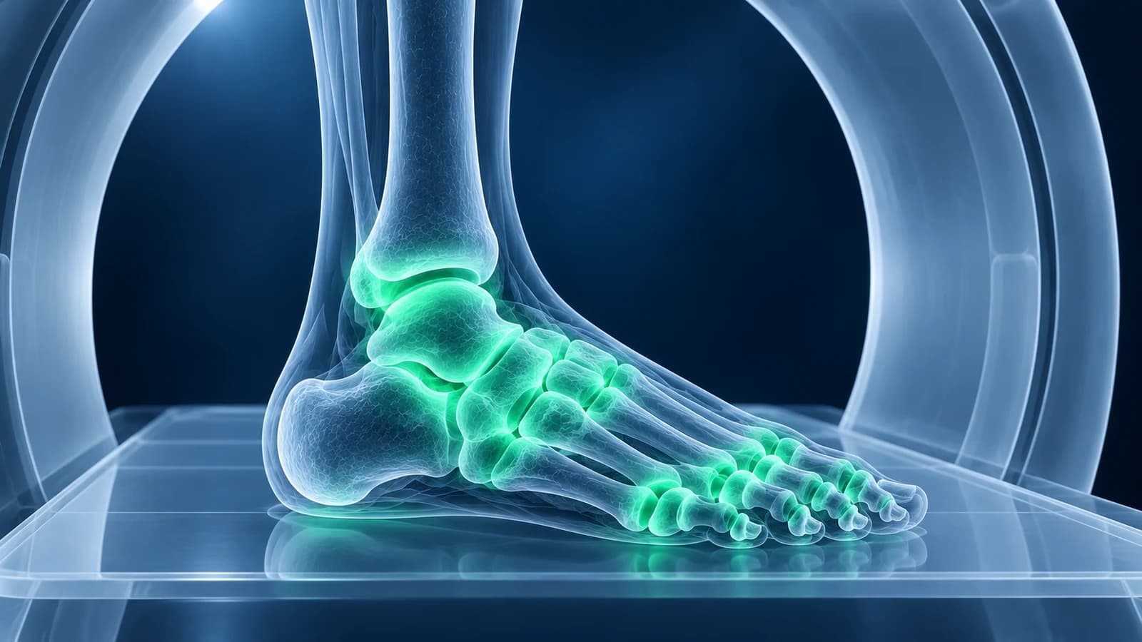
A weight-bearing CT is a 3D scan of the foot taken while you stand. Standing X-rays also show the foot under load, but only as flat pictures with overlapping bones, while MRI and ordinary CT are done lying down. Its radiation dose is lower than standard CT: a study on a model ankle measured the cone-beam CT technology most standing scanners use at roughly 1.3 to 10 times a two-view X-ray, against about 14 times for conventional CT.
This guide explains what that standing 3D view adds, which foot problems it helps with most, what it cannot show, and how to think about dose and availability. It is educational and not a recommendation for a particular test; the right scan depends on the question your clinician is trying to answer.
What is a weight-bearing CT scan?
Most weight-bearing CT scanners use cone-beam technology: a cone-shaped X-ray beam and a flat detector rotate once around the feet and the computer builds a 3D volume from the projections. A topical review in Foot & Ankle International describes how this design allows imaging of the lower limb both lying down and standing, which a conventional hospital CT, with its closed tunnel and moving table, cannot do.
In practice you step onto the platform, usually with both feet in the scanner, and stand still while it makes a single rotation. There is no tunnel, no injection and no noise of the kind MRI makes. The images can then be sliced in any direction, just like an ordinary CT, and opened as DICOM files in the free CT viewer.
What does weight-bearing CT show that standing X-rays and MRI do not?
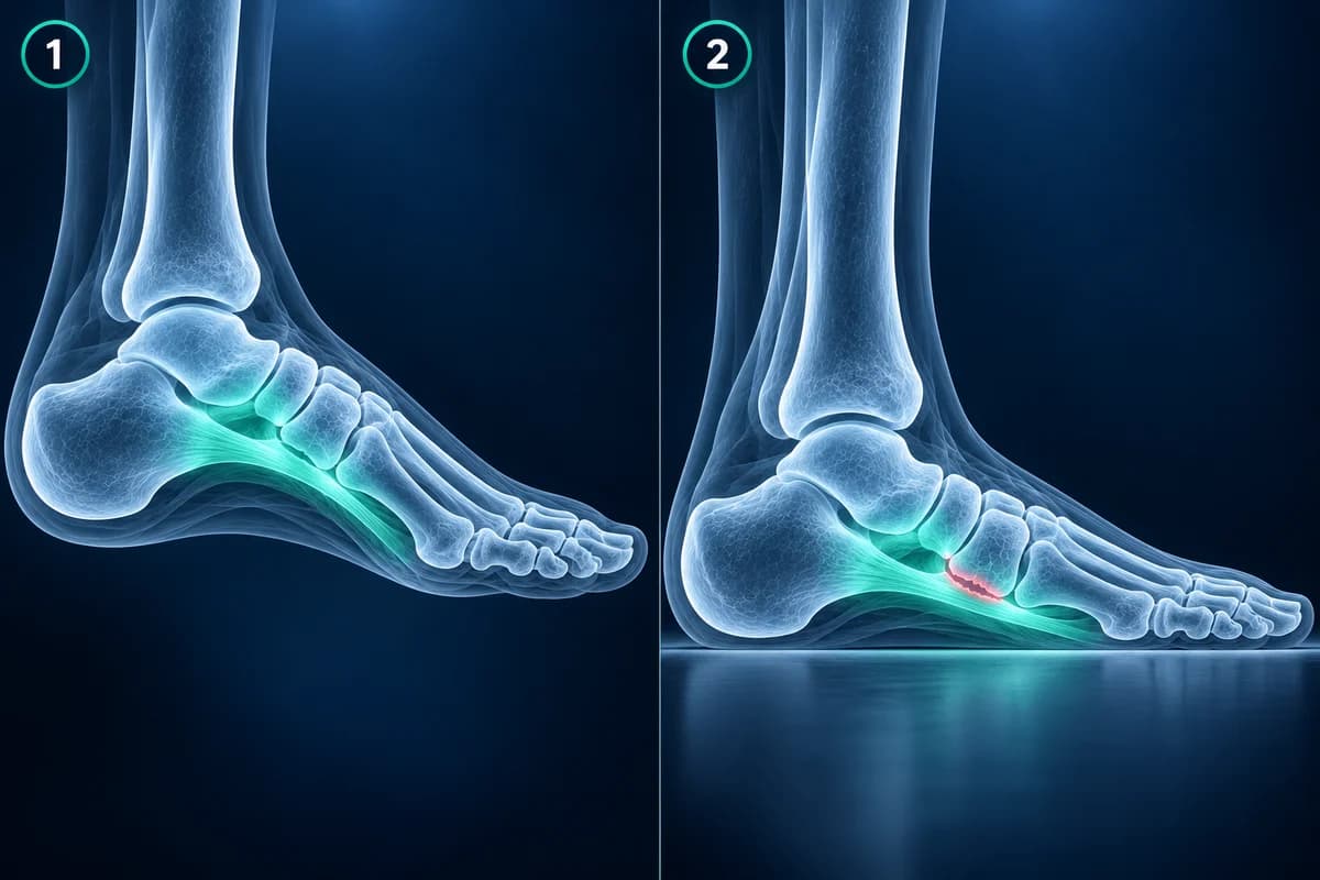
The foot is a flexible structure. When you stand, the arch lowers, the heel can tilt outward and joints that look perfectly lined up on a scan taken lying down can open, slide or press together. A standing X-ray captures that loaded position, but it squashes 26 bones into a single flat picture, so many joints overlap and angles depend on exactly how the foot was turned. MRI shows tendons, ligaments and bone marrow in great detail, yet it is done with the foot resting, so it cannot show how the bones behave under weight.
Weight-bearing CT combines the loaded position of the standing X-ray with the 3D detail of CT. Surgeons can measure alignment in all three planes, look straight into joints that hide behind other bones on X-ray, and see where bones start to touch each other when the foot collapses. The guide to foot X-ray angles explains the 2D measurements that weight-bearing CT is designed to refine.
How does weight-bearing CT help with flat foot?
Adult-acquired flat foot is now called progressive collapsing foot deformity. In 2020 an international expert group published a consensus on how to image it: standing X-rays of the foot and ankle are necessary, and if weight-bearing CT is available it is strongly recommended for planning surgery. The group listed the findings it should be checked for, including the subtalar joint slipping out of position, the heel bone pressing against the talus in the small tunnel on the outer side of the hindfoot, and the heel bone pressing up against the tip of the fibula.
Those contact points can cause the pain on the outer side of the foot that people with an advanced flat foot often describe, and they are hard to judge on a flat X-ray. The flat foot page covers the condition itself. Weight-bearing CT is also used in children and teenagers with stiff flat feet, where a tarsal coalition may be the cause.
Can weight-bearing CT find a subtle Lisfranc injury?
Possibly, and this is one of its most studied uses. A torn Lisfranc ligament can leave the midfoot looking normal when it is unloaded and only open under weight. A 2023 systematic review comparing weight-bearing CT with conventional CT concluded that the standing scan appears better at detecting subtle Lisfranc instability, likely because it assesses the joint under load. The authors also noted that more research is needed before it can be called the better test.
For now, standing X-rays of both feet remain the usual first step, and MRI is still the test that shows the ligament itself. The article on a Lisfranc injury missed on X-ray explains the X-ray signs, and the Lisfranc injury page covers treatment.
What else is weight-bearing CT used for?
Surgeons use it to plan and check operations where alignment under load matters. In a bunion, the first metatarsal often rotates as well as drifting sideways, and that rotation is easier to judge in 3D. In midfoot and hindfoot arthritis, the scan shows which joints have lost their space and whether a fusion has fully joined. Because it is CT, it also shows fracture healing well, which helps when an X-ray leaves it unclear whether a bone has bridged.
What can weight-bearing CT not show?
It is still CT, so it shows bone far better than soft tissue. It will not show a torn tendon, a plantar plate tear, a Morton's neuroma, bone marrow edema from a stress reaction, or early infection; those need MRI or ultrasound. Cone-beam images also have more noise and less soft-tissue contrast than a hospital CT, and standing still matters, because movement blurs the volume. Someone who cannot stand safely for the scan may need a conventional CT lying down instead.
If you are weighing your options, our comparison of foot MRI vs X-ray vs ultrasound explains which question each test answers best.
How much radiation does a weight-bearing CT use?
Less than a conventional CT, but more than a set of X-rays, and the exact figure depends on the machine and the settings. A dosimetry study on a model ankle in the Journal of Foot and Ankle Research measured a conventional CT at about 14 times the dose of a two-view X-ray, while two cone-beam scanners ranged from about 1.3 to 10 times depending on the field of view and mode. The foot and ankle are far from the most radiation-sensitive organs, so these doses are small in absolute terms.
In children, a study in the Journal of Pediatric Orthopaedics found an average weight-bearing CT dose of about 0.6 to 1.1 mGy, compared with about 7.9 to 10.4 mGy for conventional CT in children of similar weight. That gap is one reason the scan is increasingly used for growing feet.
Where is weight-bearing CT available?
Weight-bearing CT is still a newer, specialist test. The scanners are found mainly in foot and ankle surgery practices, some orthopedic and sports clinics and a number of university hospitals, rather than in every imaging center. Plain standing X-rays remain the first test for most foot problems, and a weight-bearing CT is usually ordered by a foot and ankle specialist who already has a specific question about alignment or a joint.
If one is suggested, it is reasonable to ask what question it will answer, how the result could change the plan, and whether your insurer covers it, because coverage varies. Prices and coverage for other foot scans are compared in our foot MRI cost guide.
Where does weight-bearing CT fit in the imaging pathway?
The ACR adult chronic-foot-pain guidance recommends radiographs first for unexplained pain. For suspected bone or joint disease with negative or unclear radiographs, CT foot without contrast or MRI without contrast is rated usually appropriate. These CT ratings are not specific to weight-bearing CT.
Frequently Asked Questions
Is a weight-bearing CT better than an MRI for the foot?
They answer different questions. Weight-bearing CT shows how the bones line up and touch under body weight. MRI shows tendons, ligaments, nerves and bone marrow edema. Alignment problems favor the standing CT; soft tissue and stress injuries favor MRI.
Why do I need a weight-bearing CT if I already had standing X-rays?
Standing X-rays show the loaded foot as a flat picture with overlapping bones. A surgeon may want the 3D view to measure alignment in all planes, see contact points in the hindfoot, or plan an operation more precisely.
How long does a weight-bearing CT take?
The scan itself is short: the scanner makes one rotation while you stand still, and the whole visit is usually brief. There is no injection, and you stay in your own clothes apart from removing shoes and socks.
Is weight-bearing CT safe for children?
It uses radiation, so it is ordered only when the answer matters. A pediatric study found its average dose was roughly a tenth of a conventional CT of the foot and ankle in children of similar weight.
Can I view my weight-bearing CT images myself?
Yes. Ask the imaging center for the DICOM files. They open in a standard CT viewer, including a browser-based viewer that keeps the files on your device, though reading 3D alignment measurements takes specialist training.
Sources
- Barg A, Bailey T, Richter M, et al. Weightbearing Computed Tomography of the Foot and Ankle: Emerging Technology Topical Review. Foot Ankle Int. 2018;39(3):376-386.
- de Cesar Netto C, Myerson MS, Day J, et al. Consensus for the Use of Weightbearing CT in the Assessment of Progressive Collapsing Foot Deformity. Foot Ankle Int. 2020;41(10):1277-1282.
- Talaski GM, Baumann AN, Walley KC, et al. Weightbearing Computed Tomography vs Conventional Tomography for Examination of Varying Degrees of Lisfranc Injuries: A Systematic Review of the Literature. Foot Ankle Orthop. 2023;8(4):24730114231209767.
- Koivisto J, Kiljunen T, Kadesjö N, et al. Effective radiation dose of a MSCT, two CBCT and one conventional radiography device in the ankle region. J Foot Ankle Res. 2015;8:8.
- Holbrook HS, Bowers AF, Mahmoud K, Kelly DM. Weight-Bearing Computed Tomography of the Foot and Ankle in the Pediatric Population. J Pediatr Orthop. 2022;42(6):321-326.
Related Articles
What the numbers on your foot X-ray mean: hallux valgus angle, intermetatarsal angle, Meary's angle, calcaneal pitch and Böhler's angle, with normal ranges and how each is measured.
X-ray, MRI, ultrasound or CT for foot pain? What each test shows and misses, which one usually comes first, and how the choice changes for stress fractures, neuromas, plantar fasciitis, Lisfranc injuries and the diabetic foot.
Why Lisfranc midfoot injuries are missed on first X-rays, the alignment lines and fleck sign to check, and when standing X-rays, CT or MRI are needed.
Related Conditions
Understand your imaging before your next appointment
MRI, X-ray or CT from a CD, folder or ZIP. A PDF report or photos of the films work too.
Free to open and preview, no account needed. An optional AI report from $10 explains your images in plain language, with questions for your doctor.
Your raw files stay on this device. Only rendered images and minimal context are sent when you request an AI report.
Medical Disclaimer: This page is for informational and educational purposes only. It does not constitute medical advice, diagnosis, or treatment. AI-generated analysis may contain errors. Always consult a qualified healthcare professional for medical decisions. Full Disclaimer