
Sever's Disease (Calcaneal Apophysitis): When a Child's Heel Needs Imaging
Sever's disease is a painful strain of the growth plate at the back of the heel in active children, most often between about 8 and 15 years old. It is diagnosed by examination, and many children never need an X-ray. A heel growth centre that looks dense or broken into pieces on X-ray is often normal, so imaging is kept for pain that does not fit the usual pattern.
Common Symptoms
- Aching at the back or sides of the heel during or after football, basketball, gymnastics or running
- Limping or walking on tiptoe at the end of a training day to keep weight off the heel
- Pain when the sides of the back of the heel are squeezed together between finger and thumb
- Heel soreness that flares at the start of a new season or during a growth spurt
- Stiffness first thing in the morning that loosens up after a few minutes of moving around
- Tight calf muscles, so the child struggles to pull the foot up toward the shin
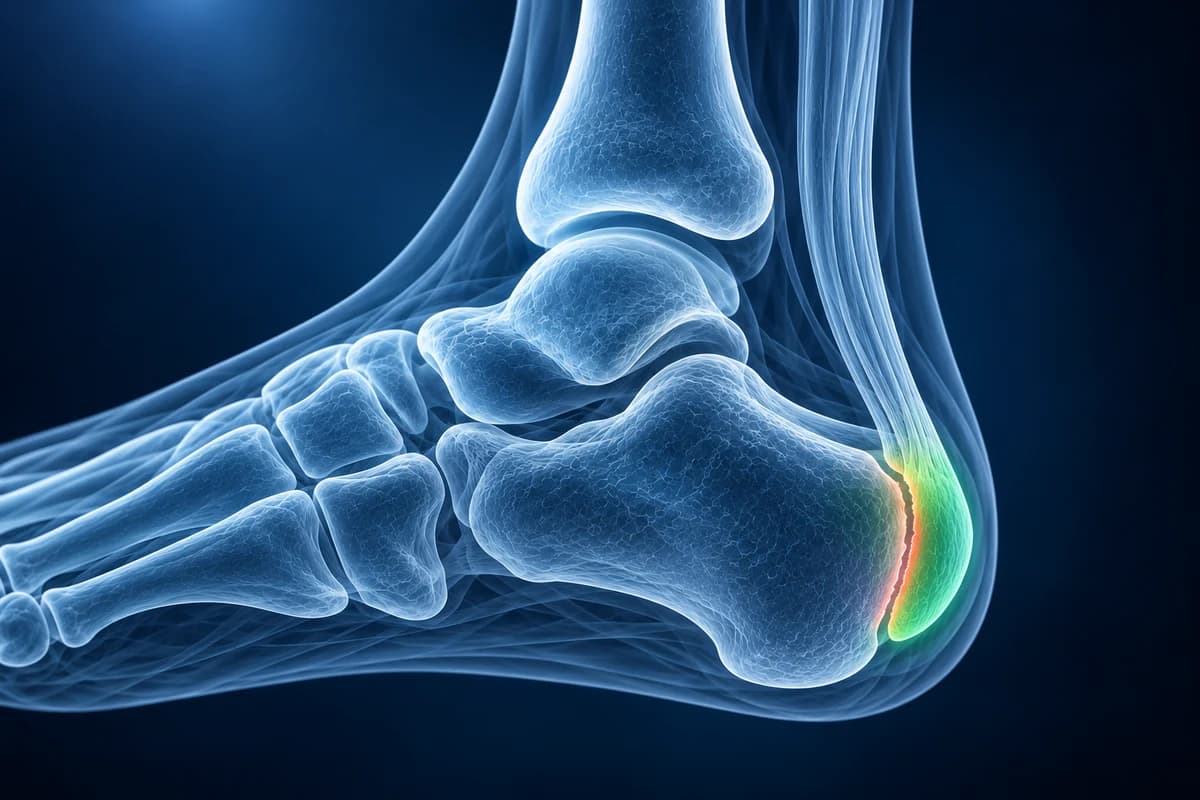
Doctors also call it calcaneal apophysitis. It is not an infection and it does not damage the bone in the long run: the pain fades as the growth plate fuses at the end of growth. This page explains when an X-ray or MRI genuinely helps, the phrases reports use, and the signs that point to something other than a growing heel. It is educational guidance, not a diagnosis, and imaging decisions for a child belong to the clinician who examines them.
When is a child's heel pain not Sever's disease?
Sever's disease hurts with activity and settles with rest. Get the child examined promptly, the same day for the first two, if any of the following apply.
- Fever, a hot red heel, or a child who seems unwell or refuses to stand
- Heel pain that began with a fall, a jump from height or a twist, and the child cannot bear weight
- Pain that wakes the child at night or is present at rest, not only during sport
- Visible swelling, a lump, or pain in one heel only that keeps worsening over weeks
- A child younger than the usual age range, weight loss, or pain in other joints as well
These features are where an X-ray, blood tests or an MRI earn their place, because bone infection, a bone cyst, a stress fracture or, rarely, a tumour can all cause heel pain in a growing child.
Does my child need an X-ray for Sever's disease?
Often not. When the story is typical (a sporty child, both heels or one heel sore after activity, pain on squeezing the back of the heel, no injury and no red flags), the diagnosis is clinical. The StatPearls review on calcaneal apophysitis states that imaging is not needed to make the diagnosis, and that X-rays are mainly useful to rule out a fracture while MRI helps separate it from infection or a tumour.
The studies disagree on how routinely to X-ray. Kose followed 61 children with the clinical diagnosis prospectively: 70 of 71 sets of foot X-rays were normal, and only one changed the diagnosis, showing a simple bone cyst. Rachel and colleagues reviewed 98 children with an average age of 10.8 years and found unexpected findings in 5.1%, including bone cysts and calcaneal stress fractures, and argued for a single side-view X-ray. In practice, many clinicians examine first and X-ray when pain is one-sided, unusually severe, present at night, or not improving after several weeks of reduced activity.
Heel pain in children has a short list of other causes, each covered in the guide to children's foot pain and imaging and, for all ages, the heel pain causes and imaging overview.
Why can a normal heel growth plate look broken on an X-ray?
The calcaneal apophysis is a separate cap of bone at the back of the heel. In Volpon's X-ray study of 392 healthy boys, it began to ossify at about 7 years and was fused in every boy by 15. On a side-view X-ray it can look whiter (sclerotic) than the rest of the heel bone and split into several pieces (fragmented). Volpon found sclerosis to be a normal feature, and fragmentation also varies in healthy children even though it was more marked in boys with heel pain. Kose likewise concluded that neither sclerosis nor fragmentation reliably makes the diagnosis.
The growth plate itself shows as a dark, wavy line between the cap and the body of the heel bone. To someone who has not seen many children's feet, that line can look like a fracture. What a radiologist checks instead is whether both heels look alike, whether the bone next to the growth plate has a hole or a lucent area, and whether the soft tissue is swollen. A plain, symmetric appearance is reassuring. You can compare both heels yourself in the free X-ray viewer; the files stay on your device.
What does “sclerosis and fragmentation of the calcaneal apophysis” mean on an X-ray report?
These are the phrases a child's heel X-ray or MRI report is most likely to contain, with a question worth bringing to the appointment for each.
- “Sclerosis and fragmentation of the calcaneal apophysis”: the heel growth cap looks dense and in pieces, which is common in healthy children and does not prove Sever's disease. Ask whether anything else on the film looked abnormal, or whether this is simply a normal growing heel.
- “Open calcaneal apophysis, no acute osseous abnormality”: the growth plate has not fused yet and no fracture was seen. Ask whether the plan is now based on the examination alone and when to come back if pain persists.
- “Lucent lesion in the calcaneus” or “unicameral bone cyst”: a fluid-filled hole in the heel bone, the most frequent unexpected finding in both X-ray studies above. Ask whether it weakens the bone enough to change sport or activity and how it will be followed.
- “Sclerotic line across the calcaneal body”: a band of denser bone that can be a healing calcaneal stress fracture rather than apophysitis. Ask whether the child needs a period of protected weight bearing.
- “Bone marrow edema in the calcaneal apophysis” on MRI: fluid in the growth cap, which fits a stressed apophysis in a sporty child but is also checked against infection. Ask whether the scan excluded osteomyelitis and a stress fracture, and whether blood tests are needed.
For the wording on adult and child foot X-rays in general, see foot X-ray report phrases explained.
When is an MRI, ultrasound or blood test used for a child's heel pain?
For chronic foot pain in general, the American College of Radiology Appropriateness Criteria place plain X-rays first and add MRI when X-rays are normal or unclear and the answer would change treatment. In a child with apophysitis-type heel pain, that usually means MRI only when red flags are present or symptoms do not follow the expected course.
MRI shows marrow swelling, a stress fracture line, bone infection, cysts and soft-tissue problems such as an inflamed bursa or irritation of the Achilles tendon insertion. It needs the child to lie still for a while, and very young children may need sedation, which is one reason it is not a routine test. A scan on disc or as DICOM files can be opened in the free MRI viewer. Ultrasound can look at the Achilles tendon and bursa without radiation but sees little of the bone. Blood tests such as inflammatory markers are added when infection or inflammatory arthritis is a concern.
What We Look For on a Child's Heel Imaging
- Symmetry of the calcaneal apophysis between the painful and the comfortable heel
- Lucent areas, cysts or bone destruction next to the growth plate
- A sclerotic band or fracture line in the body of the heel bone
- Soft-tissue swelling at the Achilles insertion or under the heel
- Marrow edema pattern on MRI and whether it is confined to the apophysis
- Clues to another diagnosis, such as a bar of bone between the tarsal bones
What else causes heel and foot pain in growing children?
A child whose foot is stiff and keeps rolling in, with repeated “ankle sprains”, may have a tarsal coalition, a bridge between two foot bones that does show on X-ray or CT. Pain on the inner side of the midfoot in a younger child, often with a limp, points toward Köhler disease or an accessory navicular rather than the heel. After a fall from height, heel pain needs an X-ray to look for a calcaneal fracture.
Pain under the heel in an adult is usually plantar fasciitis, a different condition. Sever's disease sits higher, at the back of the heel where the Achilles tendon pulls, and only exists while the growth plate is open.
How long does Sever's disease last, and how is it treated?
Sever's disease is self-limiting: symptoms come and go with sport and growth spurts and stop for good once the apophysis fuses to the heel bone. Treatment aims to keep the child active with less pain. The usual steps are reducing running and jumping to a level that does not cause a limp, ice after activity, calf stretching and strengthening, cushioned heel cups or lifts in sports shoes, and simple pain relief when needed. A short period in a walking boot or cast is reserved for severe pain.
Most children can keep playing in a reduced role, guided by pain. Stopping sport entirely is rarely necessary, and surgery is not a treatment for Sever's disease. If pain does not improve over a couple of months of sensible load management, the diagnosis is worth reviewing rather than simply resting longer.
Frequently Asked Questions
Can Sever's disease be seen on an X-ray?
Not reliably. A dense heel growth cap is normal, and a fragmented one is also seen in children without pain, so the diagnosis rests on the examination. An X-ray is mainly used to rule out a fracture, a bone cyst or another problem when the story is unusual.
At what age does Sever's disease stop?
It settles when the heel growth plate fuses, which in a large X-ray study of healthy boys had happened in every boy by age 15. Until then, flares are common at the start of a sports season or during a growth spurt and usually ease with a temporary drop in running and jumping.
Should my child stop sport with Sever's disease?
Usually not completely. Activity is adjusted so the child can play without limping, with ice, calf stretches and heel cups. A complete break is used only for severe pain. Your child's clinician or physiotherapist can set a level that suits the season.
Is Sever's disease the same as a broken heel?
No. It is irritation of a growth plate from the repeated pull of the Achilles tendon, not a break. A stress fracture of the heel bone can feel similar, which is one reason one-sided or worsening pain is X-rayed.
Can AI read my child's heel X-ray?
Sever's disease is usually diagnosed from symptoms and examination; heel X-ray changes can also occur in healthy children. AI cannot diagnose heel pain or decide whether imaging is needed.
Sources
- Smith JM, Varacallo MA. Sever Disease (Calcaneal Apophysitis). StatPearls. Treasure Island (FL): StatPearls Publishing.
- Kose O. Do we really need radiographic assessment for the diagnosis of non-specific heel pain (calcaneal apophysitis) in children? Skeletal Radiol. 2010;39(4):359-361.
- Rachel JN, Williams JB, Sawyer JR, Warner WC, Kelly DM. Is radiographic evaluation necessary in children with a clinical diagnosis of calcaneal apophysitis (Sever disease)? J Pediatr Orthop. 2011;31(5):548-550.
- Volpon JB, de Carvalho Filho G. Calcaneal apophysitis: a quantitative radiographic evaluation of the secondary ossification center. Arch Orthop Trauma Surg. 2002;122(6):338-341.
- Expert Panel on Musculoskeletal Imaging; Said N, Fox MG, Nacey NC, et al. ACR Appropriateness Criteria Chronic Foot Pain: Update 2025. J Am Coll Radiol. 2026;23(4):681-699.
Related Articles
Heel pain mapped to its causes: plantar fasciitis, heel stress fracture, Baxter's nerve, fat pad atrophy and Sever's disease, and which test shows each one.
Why a child's foot hurts: Sever's, Köhler, tarsal coalition, accessory navicular and toddler fractures, what X-rays show and the red flags to act on.
Understand your imaging before your next appointment
MRI, X-ray or CT from a CD, folder or ZIP. A PDF report or photos of the films work too.
Free to open and preview, no account needed. An optional AI report from $10 explains your images in plain language, with questions for your doctor.
Your raw files stay on this device. Only rendered images and minimal context are sent when you request an AI report.
Medical Disclaimer: This page is for informational and educational purposes only. It does not constitute medical advice, diagnosis, or treatment. AI-generated analysis may contain errors. Always consult a qualified healthcare professional for medical decisions. Full Disclaimer