
Köhler Disease: What a Flattened Navicular on a Child's X-ray Means
Köhler disease is a temporary loss of blood supply to the navicular bone in the midfoot of a young child, most often a boy aged about 4 to 7. On X-ray the navicular looks flat, dense and sometimes in pieces. It heals on its own: the bone rebuilds over months to a few years and the foot is expected to be normal in adulthood.
Common Symptoms
- A limp in a young child, often without any remembered fall or injury
- Pain and tenderness on the inner side of the midfoot, in the middle of the arch
- Mild swelling or warmth over the top and inner side of the midfoot
- Walking on the outer edge of the foot to keep weight off the arch
- Discomfort that gets worse with running, jumping or a long day on the feet
- Pain usually in one foot, although both feet are sometimes affected
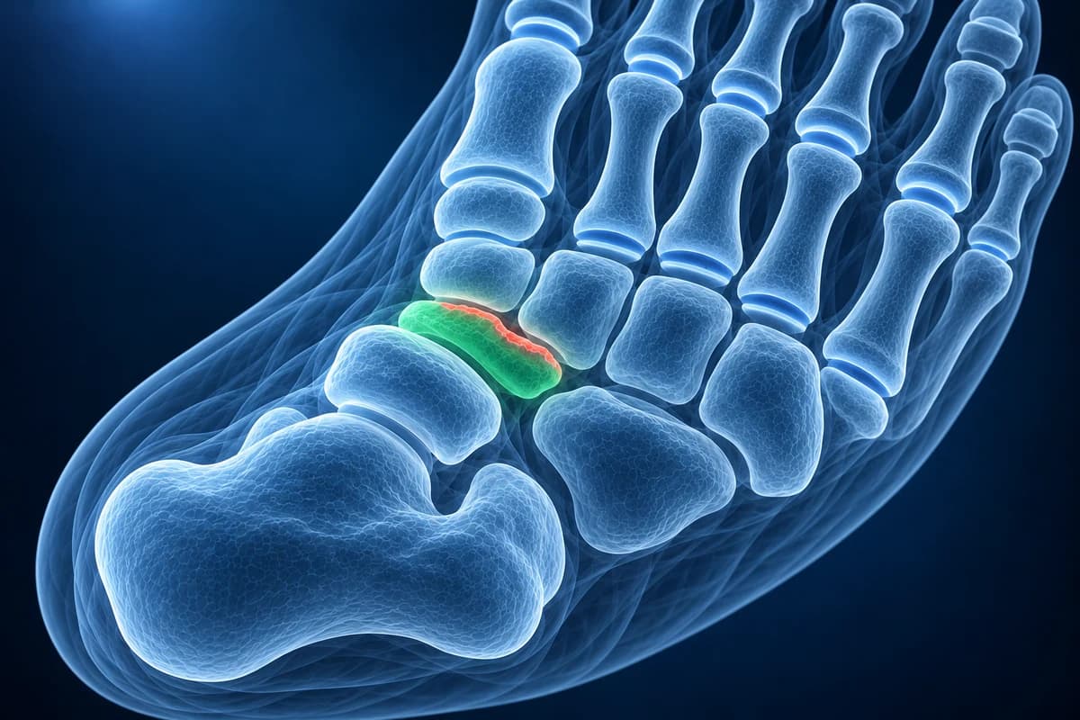
It is one of the osteochondroses, a group of conditions in which a growing bone centre temporarily loses part of its blood supply and then rebuilds. The hard part for parents is that the X-ray looks dramatic while the outlook is excellent, and that an irregular navicular can also be seen in children with no symptoms at all. This page explains how doctors tell these apart and what the report wording means. It is educational guidance, not a diagnosis, and imaging decisions for a child belong to the clinician who examines them.
When does a limping child need to be seen the same day?
- Fever, a hot and red foot, or a child who seems unwell, which can mean bone or joint infection
- Refusing to stand or walk at all, or crying when the foot is touched
- A limp after a fall or twist with swelling or bruising
- Night pain, weight loss, or pain and swelling in several joints
Köhler disease can look like infection of the navicular on an X-ray, so blood tests are often used when the child has a temperature or the foot is very swollen.
What does Köhler disease look like on an X-ray?
The diagnosis is made on plain X-rays, usually a top-down and a side view of the foot. The navicular, which sits between the ankle bone and the three wedge-shaped cuneiforms, appears smaller than expected, flattened from front to back like a wafer, and whiter (sclerotic) than its neighbours. It may also be broken into fragments. The StatPearls review describes this combination of sclerosis, fragmentation and flattening as the typical pattern of avascular necrosis, and notes that one report found both feet affected in about a quarter of cases.
The joints on either side of the navicular usually keep their normal gaps, and the other bones of the foot look healthy. That contrast, one small dense bone in an otherwise normal growing foot, is what draws the radiologist's eye. Images on disc or as DICOM files can be opened in the free X-ray viewer, and the guide to reading a foot X-ray shows where the navicular sits.
Is it Köhler disease or normal irregular ossification of the navicular?
The StatPearls review notes that the navicular is the last of the tarsal bones to turn to bone, and in some healthy children it does so unevenly: from several small centres, with a patchy or denser look for a while. On a single X-ray this can be hard to tell from Köhler disease. Williams and Cowell, reviewing 20 children with Köhler disease, made the point that true, painful Köhler disease has to be separated clinically from look-alike X-ray changes in children who have no symptoms.
In practice, the answer comes from the child more than the film. A child with no pain, no limp and no tenderness over the navicular, whose X-ray was taken for another reason, most likely has a normal variant and needs no treatment. A child with a limp and pinpoint tenderness over the same bone fits Köhler disease. Comparing with the other foot can help, but because both feet can be affected, a matching appearance does not settle the question on its own.
What does “sclerosis and flattening of the tarsal navicular” mean on a report?
Each phrase below is followed by a question that is worth asking the clinician.
- “Sclerosis, flattening and fragmentation of the tarsal navicular”: the classic Köhler pattern of a dense, thin, broken-up navicular. Ask whether the examination matches it, since the same look can occur without symptoms.
- “Irregular ossification of the navicular, may represent a normal variant”: the radiologist sees uneven bone formation but is not sure it is disease. Ask whether, given the symptoms, this needs any treatment or simply observation.
- “Findings consistent with osteochondrosis (Köhler disease)”: the X-ray fits the condition. Ask whether a short period in a walking cast or boot would help, and when a follow-up X-ray is planned.
- “Interval reconstitution of the navicular”: on a later X-ray the bone is regaining its height and normal density. Ask whether activity can now go back to normal.
- “Marrow signal abnormality in the navicular” on MRI: a change in the bone that fits loss of blood supply but can also be seen with infection or injury. Ask whether infection has been ruled out and whether blood tests were normal.
More plain-language explanations of foot X-ray wording are collected in foot X-ray report phrases explained.
Does a child with Köhler disease need an MRI?
Usually not. The American College of Radiology Appropriateness Criteria for chronic foot pain place plain X-rays first, and in a young child with typical symptoms and the classic X-ray pattern, X-rays plus a follow-up examination are generally enough. MRI is added when the picture is unclear: a high temperature, raised blood markers, swelling that is out of proportion, or symptoms that do not follow the expected course. It can show whether the change is limited to the navicular and help separate Köhler disease from bone infection or a hidden fracture, but in a small child it may need sedation.
Ultrasound adds little for the navicular itself. CT is rarely needed and uses more radiation than X-ray, which matters in children. If an MRI was done, the free MRI viewer opens it privately on your device.
What We Look For on a Child's Midfoot X-ray
- Size, height and density of the navicular compared with the neighbouring tarsal bones
- Fragmentation of the navicular and whether the pieces keep the bone's outline
- Joint spaces around the navicular and alignment of the arch
- Appearance of the navicular in the other foot, when it has been imaged
- Soft-tissue swelling, bone destruction or periosteal reaction that would suggest infection
- Extra bones beside the navicular or bony bars between tarsal bones
How long does Köhler disease take to heal?
The bone heals by itself whatever the treatment. In the series by Williams and Cowell, children who were not casted had symptoms for an average of 15 months, while those treated in a short-leg walking cast for about eight weeks had symptoms for less than three months, and every child's navicular eventually reconstituted with excellent function. Borges and colleagues followed 14 patients for an average of more than 31 years and concluded that a normal adult foot can be expected; cast treatment shortened the painful phase but did not change the final result.
Treatment is therefore about comfort: activity adjusted to pain, supportive footwear or an arch support, and a cast or boot when walking is painful. Surgery is not used. The X-ray often lags behind the child, so the bone can still look abnormal after the limp has gone.
What else causes midfoot pain in a child?
In an older child or teenager, a tender bump on the inner side of the same bone is more likely an accessory navicular, an extra bone rather than a bone losing its blood supply. A stiff flat foot with repeated sprains suggests a tarsal coalition, and heel pain in a sporty school-age child is usually Sever's disease. A similar loss of blood supply can affect the second metatarsal head in teenagers, known as Freiberg disease. The children's foot pain and imaging guide compares them side by side.
In athletes after growth has finished, pain in the same bone has a very different meaning and can be a navicular stress fracture, which needs prompt imaging.
Frequently Asked Questions
Is Köhler disease serious?
It looks worrying on X-ray but has an excellent outlook. The navicular rebuilds itself, and long-term follow-up studies report normal adult feet. The main question at diagnosis is making sure it is not an infection or an injury.
Does Köhler disease need a cast?
Not always. A short-leg walking cast for several weeks shortened the painful period in older case series, so it is often offered when the limp is marked. Milder cases are managed with activity changes and supportive shoes. Both routes end with a healed bone.
How long until the X-ray looks normal again?
Often longer than the pain lasts. The navicular can take many months or a few years to regain normal height and density, so a follow-up X-ray is timed by the clinician rather than repeated at every visit.
Can my child keep doing sport?
Usually, at a level that does not cause a limp. Running and jumping may need to be reduced during the painful phase. The treating clinician can advise when full activity is safe.
Can AI tell Köhler disease from a normal navicular?
A dense, flattened navicular on X-ray may support Köhler disease, but normal bone development can look similar and symptoms matter. AI cannot diagnose a child's limp.
Sources
- Trammell AP, Davis DD, Scott A. Kohler Disease. StatPearls. Treasure Island (FL): StatPearls Publishing.
- Williams GA, Cowell HR. Köhler's disease of the tarsal navicular. Clin Orthop Relat Res. 1981;(158):53-58.
- Borges JL, Guille JT, Bowen JR. Köhler's bone disease of the tarsal navicular. J Pediatr Orthop. 1995;15(5):596-598.
- Expert Panel on Musculoskeletal Imaging; Said N, Fox MG, Nacey NC, et al. ACR Appropriateness Criteria Chronic Foot Pain: Update 2025. J Am Coll Radiol. 2026;23(4):681-699.
Understand your imaging before your next appointment
MRI, X-ray or CT from a CD, folder or ZIP. A PDF report or photos of the films work too.
Free to open and preview, no account needed. An optional AI report from $10 explains your images in plain language, with questions for your doctor.
Your raw files stay on this device. Only rendered images and minimal context are sent when you request an AI report.
Medical Disclaimer: This page is for informational and educational purposes only. It does not constitute medical advice, diagnosis, or treatment. AI-generated analysis may contain errors. Always consult a qualified healthcare professional for medical decisions. Full Disclaimer