
Rheumatoid Arthritis of the Foot: Forefoot Erosions and What Imaging Shows
Rheumatoid arthritis often reaches the feet early, especially the joints at the base of the second to fifth toes. Foot X-rays can show erosions there, sometimes before the hands, while ultrasound and MRI detect joint lining inflammation even earlier. Imaging supports the diagnosis, but a rheumatologist makes it by combining your joints, blood tests and symptom duration.
Common Symptoms
- Pain under the ball of the foot that feels like walking on pebbles or a folded sock
- Morning stiffness in the feet that lasts longer than an hour before walking eases
- Puffy, tender swelling across the front of both feet, often matched by swollen knuckles in the hands
- Pain when the forefoot is squeezed from side to side across the toe joints
- Toes that slowly drift toward the little-toe side, curl into claws or no longer touch the ground
- Shoes that suddenly feel too tight across the front of the foot, with fatigue and a general unwell feeling
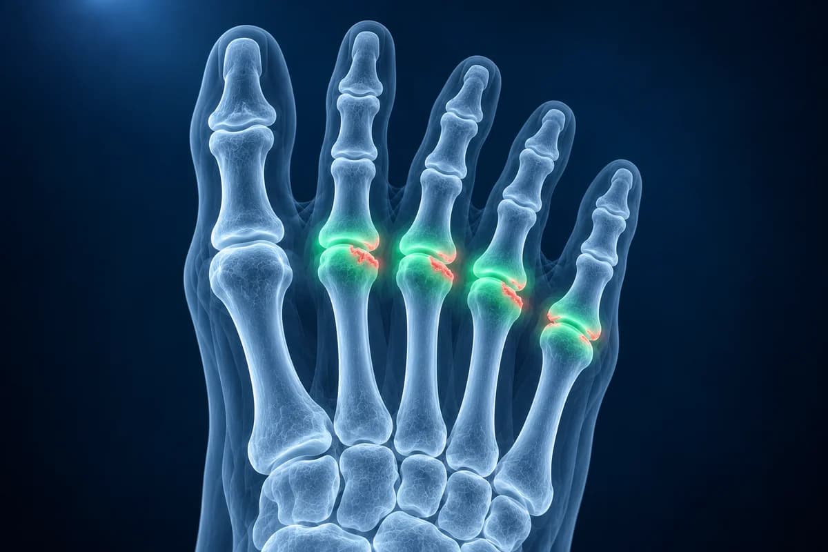
Rheumatoid arthritis is an autoimmune disease in which the lining of the joints, the synovium, becomes inflamed and thickened. Left untreated, that inflamed tissue eats into the bone at the edge of the joint and weakens the ligaments that hold the toes straight. Modern treatment started early can prevent most of that damage, which is why recognizing foot involvement matters. This page explains what each test shows, the phrases your report may use and when to seek help quickly. It is educational guidance, not a diagnosis.
Why is the forefoot often affected early in rheumatoid arthritis?
The metatarsophalangeal joints, where the toes meet the foot, are small joints lined with synovium that take the full weight of the body at every push-off. Rheumatoid arthritis targets small joints in a symmetrical pattern, and repeated loading of inflamed forefoot joints may help explain why damage often shows there first. Radiology reviews describe the fifth metatarsal head as one of the classic early sites of erosion.
That is why the international classification criteria count the second to fifth metatarsophalangeal joints among the small joints that score points, and why rheumatology guidance recommends X-rays of the feet as well as the hands when rheumatoid arthritis is suspected or diagnosed. A person may notice their feet less than their hands because forefoot pain is easily blamed on shoes or long days of standing.
Can imaging alone diagnose rheumatoid arthritis?
No. The diagnosis is made by a rheumatologist. The 2010 American College of Rheumatology and European League Against Rheumatism classification criteria add up points for the number and size of swollen joints, blood tests for rheumatoid factor and anti-CCP antibodies, inflammation markers such as CRP or ESR, and symptoms lasting at least six weeks. A score of 6 or more out of 10 classifies definite rheumatoid arthritis.
Imaging plays a supporting role. Ultrasound or MRI can confirm that a joint is genuinely inflamed when the examination is unclear, and erosions typical of rheumatoid arthritis carry strong weight. But erosions, joint fluid and bone edema also occur in other diseases, so a report alone cannot settle the diagnosis. National guidance from NICE advises referral to a specialist for suspected persistent joint lining inflammation of unknown cause, and urgent referral, even when blood tests are normal, when the small joints of the hands or feet are involved, more than one joint is affected, or there has been a delay of three months or longer between the first symptoms and seeking medical advice.
What do X-ray, ultrasound and MRI each show in the rheumatoid foot?
X-ray is the starting point. European League Against Rheumatism imaging recommendations name plain X-rays of the hands and feet as the first test for joint damage, and NICE advises X-rays of the hands and feet both when rheumatoid arthritis is suspected and soon after diagnosis, to look for erosions and give a baseline for later comparison. In early disease the X-ray is often normal, which does not rule rheumatoid arthritis out. Your images can be opened in the free X-ray viewer.
Ultrasound shows thickened synovium, joint fluid and blood flow in the inflamed lining on power Doppler, and it finds small erosions that X-ray misses. It is quick, radiation-free and can scan many toe joints in one visit, and it can guide a steroid injection into a single stubborn joint.
MRI shows synovitis, tendon sheath inflammation and bone marrow edema, which the same recommendations describe as a strong predictor of future erosions in early disease. It is used when the picture is unclear rather than as a routine test. An MRI you received as DICOM files can be opened in the free MRI viewer, and the comparison of foot MRI, X-ray and ultrasound lays out the trade-offs.
What does “marginal erosions at the metatarsal heads” mean on your report?
These are the phrases foot imaging reports use most often in inflammatory arthritis. The foot MRI report phrases guide covers the wider vocabulary.
- “Marginal erosions at the second and fifth metatarsal heads”: small bites of bone missing at the edge of the joint, where inflamed lining touches bone not covered by cartilage. Ask whether the pattern fits rheumatoid arthritis and whether it changes treatment.
- “Periarticular osteopenia”: the bone next to the joints looks thinner on X-ray, an early and nonspecific sign of inflammation. Ask whether a follow-up X-ray is planned as a baseline.
- “Uniform joint space narrowing”: cartilage has thinned evenly across the joint, typical of inflammatory arthritis rather than wear, which tends to narrow one side. Ask how it compares with earlier images.
- “Synovitis with power Doppler signal”: the joint lining is thickened and has increased blood flow, meaning active inflammation now. Ask whether it means your treatment needs adjusting.
- “Bone marrow edema (osteitis) in the metatarsal heads” on MRI: inflammation inside the bone, linked to a higher risk of later erosions. Ask how it affects the urgency of treatment.
- “Intermetatarsal bursitis”: a fluid-filled sac between the metatarsal heads, common in rheumatoid arthritis and easily confused with a nerve problem. Ask whether it explains numbness or sharp pain between the toes.
What We Look For on Rheumatoid Foot Imaging
- Marginal erosions at the metatarsal heads, especially the fifth, and at the base of the big toe
- Periarticular osteopenia and uniform joint space narrowing in a symmetrical pattern across both feet
- Synovial thickening, joint fluid and power Doppler signal on ultrasound
- Bone marrow edema and tenosynovitis on MRI
- Toe drift, partial dislocation of the toe joints and downward displacement of the metatarsal heads
- Intermetatarsal bursitis, rheumatoid nodules and changes in the hindfoot or ankle
How does rheumatoid arthritis change the shape of the forefoot?
Long periods of joint inflammation stretch the capsule and the plantar plate under each toe joint. The toes then drift toward the little-toe side, rise up off the ground and curl into hammer or claw toes. The fat pad slides forward, leaving the metatarsal heads pushed down with little cushioning, which causes the painful calluses of metatarsalgia. The big toe often turns outward into a bunion at the same time.
Severe deformity has become less common since early treatment with disease-modifying drugs became standard. Podiatry, cushioned insoles, wide and deep shoes and, when needed, forefoot reconstruction surgery help people whose feet have already changed shape. The guide to ball of foot pain causes compares rheumatoid forefoot pain with other common causes.
What else can look like rheumatoid arthritis in the foot?
Gout usually strikes one joint suddenly, most often the big toe, and its erosions have an overhanging edge. Psoriatic arthritis can swell a whole toe like a sausage and adds new bone next to erosions. Osteoarthritis narrows joints unevenly and forms spurs without erosions. A Morton's neuroma causes burning between two toes and can coexist with rheumatoid bursitis in the same space.
When is a rheumatoid foot problem urgent?
- One joint becomes suddenly hot, very painful and swollen, especially with fever or while taking immune-suppressing medicines. A joint infection needs same-day assessment.
- A sore or ulcer over a toe joint or under a metatarsal head, particularly if it is red, leaking or smells.
- New swelling in several joints of the hands or feet that has not been assessed yet. Early specialist review protects the joints.
- Numbness, color change or coldness in the toes.
Frequently Asked Questions
Can rheumatoid arthritis start in the feet?
Yes. The joints at the base of the toes are among the most common early sites, and some people have erosions in the feet before any appear in the hands. Forefoot pain with morning stiffness and swelling in both feet is worth discussing with a doctor, even if the hands feel fine.
Can a normal foot X-ray rule out rheumatoid arthritis?
No. Erosions take time to become visible on X-ray, so early rheumatoid arthritis often has a normal X-ray. The diagnosis rests on the examination and blood tests, and ultrasound or MRI can show inflammation that the X-ray cannot.
Is ultrasound or MRI better for rheumatoid arthritis in the foot?
Both detect inflammation better than examination alone. Ultrasound is quicker, cheaper and can check many joints and guide injections. MRI also shows bone marrow edema, which helps predict future damage. Which one is used depends on the question and on local practice.
What does erosive disease mean on a foot X-ray?
It means the radiologist sees bone loss at the joint edges caused by inflammation. In rheumatoid arthritis, erosions signal active disease that has already caused damage, so rheumatologists use them to judge how strongly to treat. Ask whether the erosions are new compared with earlier images.
Can AI read my rheumatoid foot imaging report?
Erosions and synovitis may support rheumatoid arthritis, but diagnosis combines symptoms, joint examination and blood tests. AI cannot diagnose rheumatoid arthritis from imaging alone.
Sources
- Aletaha D, Neogi T, Silman AJ, et al. 2010 rheumatoid arthritis classification criteria: an American College of Rheumatology/European League Against Rheumatism collaborative initiative. Ann Rheum Dis. 2010;69(9):1580-1588.
- Colebatch AN, Edwards CJ, Østergaard M, et al. EULAR recommendations for the use of imaging of the joints in the clinical management of rheumatoid arthritis. Ann Rheum Dis. 2013;72(6):804-814.
- Jacobson JA, Girish G, Jiang Y, Resnick D. Radiographic evaluation of arthritis: inflammatory conditions. Radiology. 2008;248(2):378-389.
- National Institute for Health and Care Excellence. Rheumatoid arthritis in adults: management (NG100).
- Expert Panel on Musculoskeletal Imaging; Said N, Fox MG, Nacey NC, et al. ACR Appropriateness Criteria Chronic Foot Pain: Update 2025. J Am Coll Radiol. 2026;23(4):681-699.
Understand your imaging before your next appointment
MRI, X-ray or CT from a CD, folder or ZIP. A PDF report or photos of the films work too.
Free to open and preview, no account needed. An optional AI report from $10 explains your images in plain language, with questions for your doctor.
Your raw files stay on this device. Only rendered images and minimal context are sent when you request an AI report.
Medical Disclaimer: This page is for informational and educational purposes only. It does not constitute medical advice, diagnosis, or treatment. AI-generated analysis may contain errors. Always consult a qualified healthcare professional for medical decisions. Full Disclaimer