
Toe Fracture: X-ray, Buddy Taping and When a Broken Toe Needs More
Most broken lesser toes are treated with buddy taping to the next toe and a stiff-soled shoe, usually for 3 to 6 weeks. An X-ray matters most when the toe looks bent or twisted, when the big toe is injured, when there is a wound, or when you cannot walk. The big toe is the exception: it carries push-off, so displaced fractures there may need more.
Common Symptoms
- Throbbing pain in a toe right after stubbing it, kicking something hard or dropping a weight on it
- Swelling and bruising that spreads along the toe and sometimes onto the top of the forefoot
- Pain when walking, especially at push-off or in a tight or flexible shoe
- A toe that looks bent, twisted or out of line with its neighbours
- Blood collecting under the toenail after a crush injury
- Tenderness directly over one small bone of the toe rather than at a joint
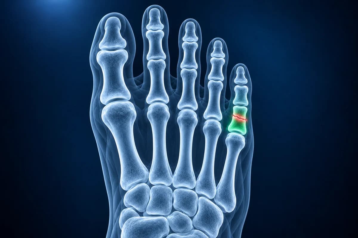
Toe fractures are among the most common fractures seen in primary care, usually from a stubbed toe or something heavy landing on the foot. In one large hospital series the big toe and the little toe were the two broken most often. This page explains when an X-ray is worth having, what the report wording means and which findings change the plan. It is general information, not a diagnosis.
Do you need an X-ray for a broken toe?
Not always. There is little research on exactly when a toe needs an X-ray, and a lesser toe that is swollen and bruised but straight, with pain you can walk on, is treated with buddy taping and a stiff shoe whether or not a hairline crack shows. Hatch and Hacking, writing for family physicians, list the situations that change treatment: a toe that is visibly deformed, a fracture of the big toe, a wound over the injury, a dislocation, a crack into a joint, or poor circulation in the toe. These are the cases where an X-ray matters most.
The Ottawa foot rules, often quoted after a foot injury, were built for the midfoot, not the toes. In their validated form they recommend an X-ray when there is pain in the midfoot together with tenderness over the navicular bone on the inner side or the base of the fifth metatarsal on the outer side, or when you could not take four steps both right after the injury and when examined. A pain mainly on the outer edge of the foot belongs with the Jones fracture page rather than here. How these rules separate a fracture from a sprain is explained in the guide to broken foot or sprain and the X-ray rules. For pain extending beyond the injured toe, compare the locations in the forefoot pain guide.
Which imaging shows a toe fracture best?
For an injured foot, the American College of Radiology Appropriateness Criteria for acute foot trauma start with X-rays. For a toe, Hatch and Hacking describe three views: top-down, side-on and oblique. Neighbouring toes overlap on the side view and can hide a crack, so the top-down and oblique views are often the easiest to read, and the oblique view can show a spiral crack the side view misses.
- X-ray shows the fracture line, whether pieces have shifted or rotated, and whether the crack runs into a joint. It is all most toe injuries ever need.
- CT is occasionally used for a complex big toe fracture into the joint, to measure how much of the surface is involved before deciding on surgery.
- MRI is rarely used for a fresh injury. It becomes useful when pain continues for weeks with a normal X-ray, to look for a hidden stress fracture, a sesamoid problem or a ligament injury under the big toe.
If you have your X-ray files, the free foot X-ray viewer lets you zoom in on each toe and switch between the views.
What does “nondisplaced fracture of the proximal phalanx” mean on your X-ray report?
Toe X-ray reports name the toe, the bone and the shape of the break. These are the phrases that come up most, with a question worth asking for each.
- “Nondisplaced fracture of the proximal phalanx of the fifth toe”: a crack in the bone nearest the foot, with the pieces still lined up. Ask whether buddy taping and a stiff shoe are enough and when to return to normal shoes.
- “Displaced”, “angulated” or “rotated”: the pieces have shifted, bent or twisted. Ask whether the toe needs to be straightened under local anaesthetic and whether a repeat X-ray will check that it stays in place.
- “Intra-articular extension”: the crack reaches a joint surface. Ask how much of the joint is involved, because a larger joint fragment, especially in the big toe, can need fixation to protect the joint.
- “Comminuted tuft fracture of the distal phalanx”: the tip bone is broken into several small pieces, typical of a crush. Ask whether the nail bed or skin is injured, since a wound that reaches a broken bone is treated as an open fracture because of the infection risk.
- “Salter-Harris fracture of the base of the proximal phalanx”: in a child, the break runs through the growth plate. Ask whether the growth plate is aligned and whether follow-up X-rays are planned.
- “No acute osseous abnormality”: no fracture was seen. Ask whether a sprain is likely and what would prompt a repeat X-ray if pain does not settle within a couple of weeks.
Other wording from foot X-rays, such as “cortical irregularity” or “accessory ossicle”, is translated in foot X-ray report phrases explained.
What We Look For on a Toe X-ray
- Which toe and which phalanx is broken, and where along the bone the line runs
- Displacement, angulation or rotation of the fracture pieces
- Extension of the fracture into the joint and the share of the joint surface involved
- Dislocation of a toe joint alongside or instead of a fracture
- Growth plate involvement in children and teenagers
- Injury to the sesamoids under the big toe or the metatarsal heads behind the toes
Can a broken toe be buddy taped, or does it need to be set?
A lesser toe fracture that is in line is usually buddy taped: a strip of gauze between the injured toe and its neighbour, then tape around both, so the healthy toe acts as a splint. A stiff-soled shoe or post-operative shoe limits bending while you walk. The American Academy of Orthopaedic Surgeons notes it can take 6 to 8 weeks before normal shoes feel comfortable, and some ache and swelling can last for 3 to 6 months.
A lesser toe that is clearly bent or twisted is usually straightened by a clinician after numbing the toe, then taped. Referral to a specialist is mainly for fractures that will not stay in place once straightened, fracture-dislocations, open injuries or poor circulation.
Why is a broken big toe treated differently?
The big toe carries much of the body's weight as you push off, so a poor result there affects walking far more than a stiff little toe would. Hatch and Hacking recommend referral to a specialist for big toe fractures that are unstable or that involve more than 25 percent of the joint surface, and note that displaced big toe fractures often need referral to keep them in place. A stable, undisplaced big toe fracture is usually buddy taped with a rigid-soled shoe for at least 4 weeks, sometimes with a walking cast.
A fracture that runs into the big toe joint can damage the cartilage and lead to stiffness and arthritis years later, the problem described on the hallux rigidus page. A forced upward bend of the big toe without a fracture can instead tear the ligaments under the joint, which is turf toe.
When does a toe injury need same-day care?
- There is a cut over the broken toe, a torn nail bed or bone visible through the skin.
- The toe is visibly bent, twisted or dislocated.
- The toe turns pale, blue, cold or numb.
- You cannot put weight on the foot, or swelling and bruising spread across the top or sole of the midfoot.
- You have diabetes or poor circulation, where taping and swelling can break the skin and a small wound can become infected.
An AI explanation can help you prepare questions for your clinician. It cannot confirm an injury or its grade on its own and is not a diagnosis.
Frequently Asked Questions
How long does a broken toe take to heal?
Most lesser toe fractures are buddy taped for 3 to 6 weeks, until the bone is no longer tender. Big toe fractures and fractures into a joint can take longer, and ache and swelling can last for a few months. The metatarsal fracture healing timeline covers the longer bones just behind the toes.
Can you walk on a broken toe?
Usually yes, as pain allows, in a stiff-soled shoe that stops the toe from bending. Being unable to take weight at all is a reason to have the foot examined, because it can mean a more serious injury further back in the foot.
Why did my X-ray miss a broken toe?
Small hairline cracks can be hidden by overlapping toes or only show on one view, and a stress fracture may not be visible for the first 2 to 4 weeks. If pain stays focused on one bone, a repeat X-ray or an MRI can show it.
Is a broken toe ever an emergency?
It is when the skin is broken over the fracture, the toe is badly deformed, or the toe loses its colour or feeling. These need same-day care because of infection risk or damage to blood vessels and nerves.
Can AI read my toe X-ray?
Toe X-rays show fracture position, alignment and joint involvement, while examination checks skin, feeling and circulation. AI cannot determine whether reduction or urgent treatment is needed.
Sources
- Hatch RL, Hacking S. Evaluation and management of toe fractures. Am Fam Physician. 2003;68(12):2413-2418.
- Expert Panel on Musculoskeletal Imaging; Gorbachova T, Chang EY, Ha AS, et al. ACR Appropriateness Criteria Acute Trauma to the Foot. J Am Coll Radiol. 2020;17(5S):S2-S11.
- Stiell IG, Greenberg GH, McKnight RD, et al. Decision rules for the use of radiography in acute ankle injuries: refinement and prospective validation. JAMA. 1993;269(9):1127-1132.
- Van Vliet-Koppert ST, Cakir H, Van Lieshout EM, et al. Demographics and functional outcome of toe fractures. J Foot Ankle Surg. 2011;50(3):307-310.
- American Academy of Orthopaedic Surgeons. Toe and Forefoot Fractures. OrthoInfo.
Understand your imaging before your next appointment
MRI, X-ray or CT from a CD, folder or ZIP. A PDF report or photos of the films work too.
Free to open and preview, no account needed. An optional AI report from $10 explains your images in plain language, with questions for your doctor.
Your raw files stay on this device. Only rendered images and minimal context are sent when you request an AI report.
Medical Disclaimer: This page is for informational and educational purposes only. It does not constitute medical advice, diagnosis, or treatment. AI-generated analysis may contain errors. Always consult a qualified healthcare professional for medical decisions. Full Disclaimer