
Broken Foot or Sprain? Ottawa Foot Rules and When to X-Ray
When a foot injury needs an X-ray: the Ottawa foot rules in plain words, the three standard views, and why a suspected Lisfranc injury needs standing films.
Key Takeaways
- Swelling and bruising cannot tell a broken foot from a sprain on their own
- The Ottawa foot rules recommend an X-ray for midfoot pain with bone tenderness at the fifth metatarsal base or navicular, or inability to take four steps
- A systematic review found the rules have a sensitivity of almost 100% for midfoot fractures
- A standard foot series has three views: front, oblique and side
- A suspected Lisfranc injury needs standing X-rays, ideally compared with the other foot
- A normal first X-ray with ongoing pain can be followed by repeat films, CT or MRI
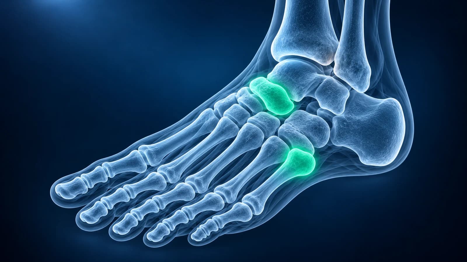
You cannot reliably tell a broken foot from a sprain by how swollen or bruised it looks. Clinicians use the Ottawa foot rules instead: an X-ray is needed when midfoot pain comes with tenderness on the bone at the base of the fifth metatarsal or at the navicular, or when you could not take four steps. A systematic review found the rules miss almost no midfoot fractures.
This guide puts the rules into plain words, explains the three X-ray views a foot series uses, and covers the one common injury the rules are not built to catch on their own: a Lisfranc injury, which often needs standing X-rays. It is educational and not a diagnosis. If you are unsure whether your foot is broken, get it examined.
What are the Ottawa foot rules in plain words?
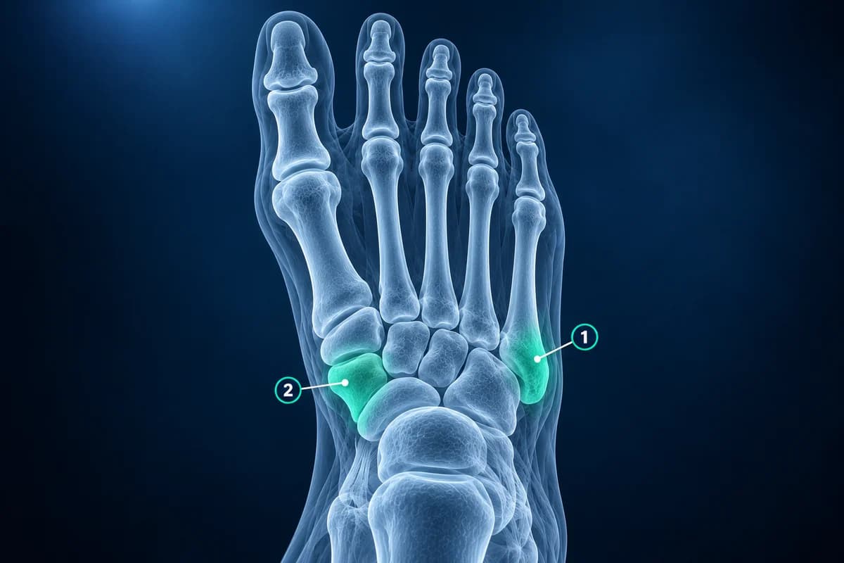
The Ottawa rules were developed in Canadian emergency departments in the early 1990s to decide which ankle and foot injuries really need an X-ray. The foot part applies when the pain sits in the midfoot, the middle section between the ankle and the base of the toes. In that case a foot X-ray series is recommended if at least one of these is true:
- Pressing on the bony bump on the outer edge of the foot, halfway along, hurts. That bump is the base of the fifth metatarsal.
- Pressing on the bony bump on the inner side of the midfoot, just in front of the ankle, hurts. That bump is the navicular.
- You could not take four steps on the foot right after the injury, and still cannot when you are examined, even if you limp.
Tenderness means pain when a finger presses directly on the bone, not general soreness of the swollen area. Pain and bruising around the outer ankle bones fall under the separate ankle part of the rules, which the ankle sprain guide on Analyze My Ankle explains.
How accurate are the Ottawa foot rules?
They are designed to be very sensitive, so that a negative result safely rules a fracture out. A systematic review in the BMJ pooled the validation studies and found a sensitivity of almost 100% for ankle and midfoot fractures, with modest specificity. In practice that means many people who meet the rules turn out to have only a sprain, but very few people who do not meet them have a fracture. The same review estimated that using the rules cuts unnecessary X-rays by about 30 to 40%.
The rules only work when the exam is reliable. They were developed in adults and have since been shown to work in children older than 5. The American College of Radiology lists situations where they should not be used, or only with great caution: reduced feeling in the foot, such as diabetic nerve damage, where the foot should simply be X-rayed; reduced alertness, for example from alcohol or a head injury; several injuries at once; a penetrating injury or any skin wound; pregnancy; a known bone disease; and an injury more than 10 days old or a return visit for ongoing pain. A clinician may also skip the rules and image directly when the foot is visibly deformed or the injury was high energy.
Which X-ray views does a foot series include?
The American College of Radiology recommends a three-view foot series as the first test after acute foot trauma when imaging is needed. The front view, taken from above and often labeled AP or dorsoplantar, shows the toes, metatarsals and the line of the inner tarsometatarsal joints. The oblique view tilts the foot inward so the outer metatarsals and the cuboid stop overlapping; it is usually the best view for the base of the fifth metatarsal. The side view, or lateral, shows the heel bone, the talus and the height of the arch.
Each view hides something the others show, which is why a single picture is not enough. A hairline crack can be invisible on two views and clear on the third. If the pain is mainly in the heel after a fall from height, extra heel views or CT are often added. The guide to how to read a foot X-ray walks through each view, and you can scroll through your own series in the free X-ray viewer, which opens the files in your browser without uploading them.
What does a fracture at each Ottawa point usually mean?
At the base of the fifth metatarsal, an inward roll of the foot often pulls off a small flake of bone where a tendon attaches. According to the AAOS, most of these avulsion fractures are treated with a walking boot or cast and weight bearing as tolerated. A break a little further along the bone, at the junction of its base and shaft, is a Jones fracture. It sits in an area with a poorer blood supply, heals more slowly and may not heal at all, so surgery is considered more often, especially in athletes. The exact position on the oblique view decides which one it is.
At the navicular, a sudden injury can chip the edge of the bone, but navicular pain that builds up during running or jumping without a single injury points to a navicular stress fracture. That is a different problem: stress fractures are often invisible on the first X-ray, and the rules for an acute twist do not apply to them.
Why does a suspected Lisfranc injury need weight-bearing X-rays?
The Lisfranc joints link the metatarsals to the midfoot. When only the ligaments tear, the bones can sit in a normal position while you lie or sit for the X-ray, then separate when you stand. The ACR criteria for acute foot trauma therefore advise that, when a Lisfranc injury is suspected, the front view be taken standing if you can bear weight, ideally with the other foot for comparison. A widened gap between the first and second metatarsal bases compared with the healthy side is the key finding.
Warning signs are pain and swelling across the top of the midfoot, pain that gets worse when you stand, walk or push off, and bruising on the sole of the foot, which the AAOS describes as highly suggestive of a Lisfranc injury. A foot that was called a sprain but still hurts to stand on or push off deserves a second look. Our article on a Lisfranc injury missed on X-ray covers the alignment lines in detail, and the Lisfranc injury page explains treatment.
What if the X-ray is normal but the foot still hurts?
A normal first X-ray does not end the story when pain persists. According to the ACR, when the X-rays are negative but a fracture or significant ligament injury is still suspected, CT or MRI may be appropriate next. CT shows small fractures and joint surfaces in detail. MRI shows bone bruising, hidden and stress fractures, and ligament tears, which is why it is chosen when the question is about soft tissue or a fracture that has not shown on X-ray.
Some clinicians simply protect the foot for a week or two and repeat the X-ray, because a crack can become visible as healing begins. The article on a stress fracture that does not show on X-ray explains why early films can look normal.
What about a stubbed or broken toe?
The Ottawa rules cover the ankle and the midfoot, not the toes. The AAOS notes that most broken toes heal without surgery, often with the toe taped to its neighbor and a wider shoe while walking is painful, so a bruised toe that is still straight is often managed without imaging. An X-ray becomes more useful when the toe is visibly bent out of line, the big toe is involved, there is a wound over the injury, or the pain is not easing after a few weeks. The toe fracture page covers the patterns that need more than strapping.
When should a foot injury be seen urgently?
Go to emergency care straight away if the foot or toes are clearly deformed, bone shows through a wound, the toes turn pale, blue, cold or numb, or the pain is severe and keeps rising with tense swelling despite rest and elevation. If you have diabetes or reduced feeling in your feet, have any foot injury checked promptly, because the usual warning pain may be missing.
Frequently Asked Questions
How can I tell if my foot is broken or just sprained?
Not reliably on your own. A clinician presses on the base of the fifth metatarsal and the navicular and checks whether you can take four steps. Tenderness at either point or inability to walk means an X-ray is advised; otherwise a fracture is unlikely.
Can you walk on a broken foot?
Sometimes, yes. Small avulsion fractures and some metatarsal fractures still allow a painful limp. Being able to walk does not rule out a fracture; bone tenderness at the Ottawa points still calls for an X-ray.
Do the Ottawa foot rules apply to children?
The original rules were developed in adults. A pooled analysis of studies in children found they reliably rule out ankle and midfoot fractures in children older than 5, missing very few. In younger children the exam is less reliable, so clinicians use more judgement. A limping child with a painful foot should be examined.
Why was my X-ray taken standing up?
Standing views load the foot, which can open a gap at the Lisfranc joints that stays hidden when the foot is resting. They are recommended when a midfoot ligament injury is suspected and you can bear weight.
My X-ray was normal. Do I need an MRI?
Not usually straight away. Most foot sprains with a normal X-ray improve over a few weeks. If pain, swelling or difficulty pushing off persists, ask whether a repeat X-ray, CT or MRI is needed to look for a hidden fracture or ligament injury.
Sources
- Expert Panel on Musculoskeletal Imaging; Gorbachova T, Chang EY, Ha AS, et al. ACR Appropriateness Criteria Acute Trauma to the Foot. J Am Coll Radiol. 2020;17(5S):S2-S11.
- Stiell IG, Greenberg GH, McKnight RD, et al. Decision rules for the use of radiography in acute ankle injuries. Refinement and prospective validation. JAMA. 1993;269(9):1127-1132.
- Bachmann LM, Kolb E, Koller MT, et al. Accuracy of Ottawa ankle rules to exclude fractures of the ankle and mid-foot: systematic review. BMJ. 2003;326(7386):417.
- Dowling S, Spooner CH, Liang Y, et al. Accuracy of Ottawa Ankle Rules to exclude fractures of the ankle and midfoot in children: a meta-analysis. Acad Emerg Med. 2009;16(4):277-287.
- American Academy of Orthopaedic Surgeons. Toe and Forefoot Fractures. OrthoInfo.
- American Academy of Orthopaedic Surgeons. Lisfranc (Midfoot) Injury. OrthoInfo.
Related Articles
Why Lisfranc midfoot injuries are missed on first X-rays, the alignment lines and fleck sign to check, and when standing X-rays, CT or MRI are needed.
A plain-language guide to your foot X-ray: the three standard views, how to trace alignment from heel to toes, and what fractures, fleck signs, accessory bones and arthritis look like.
Why a foot stress fracture can hide on a normal X-ray, what MRI, CT and repeat X-rays show instead, and which foot stress fractures are high risk.
Related Conditions
Understand your imaging before your next appointment
MRI, X-ray or CT from a CD, folder or ZIP. A PDF report or photos of the films work too.
Free to open and preview, no account needed. An optional AI report from $10 explains your images in plain language, with questions for your doctor.
Your raw files stay on this device. Only rendered images and minimal context are sent when you request an AI report.
Medical Disclaimer: This page is for informational and educational purposes only. It does not constitute medical advice, diagnosis, or treatment. AI-generated analysis may contain errors. Always consult a qualified healthcare professional for medical decisions. Full Disclaimer